

Localization and orientation of the gamma-tubulin small complex components using protein tags as labels for single particle EM
- PMID: 19723581
- PMCID: PMC2793330
- DOI: 10.1016/j.jsb.2009.08.012
Localization and orientation of the gamma-tubulin small complex components using protein tags as labels for single particle EM
Abstract
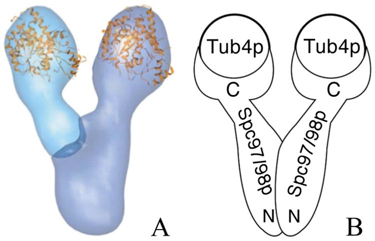
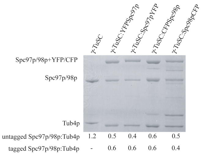
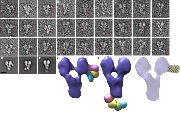
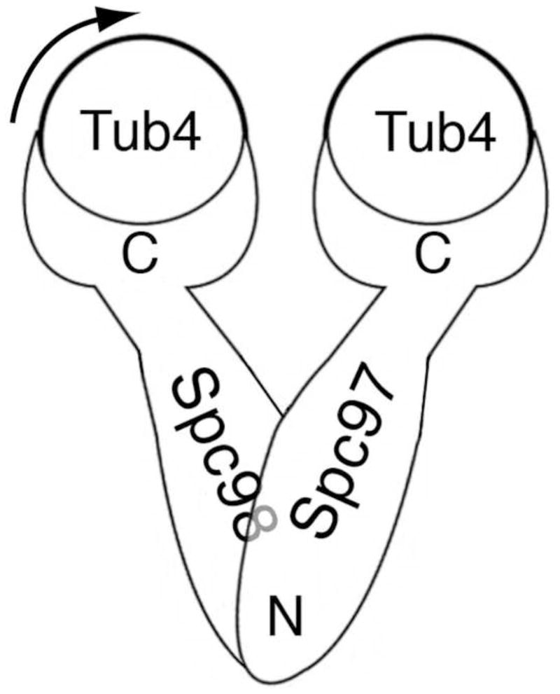
Gamma-Tubulin Small Complex (gamma-TuSC) is the universally-conserved complex in eukaryotes that contains the microtubule (MT) nucleating protein: gamma-tubulin. gamma-TuSC is a heterotetramer with two copies of gamma-tubulin and one copy each of Spc98p and Spc97p. Previously, the structure of gamma-TuSC was determined by single particle electron microscopy (EM) at 25A resolution. gamma-TuSC is Y-shaped with a single flexible arm that could be the key to regulating MT nucleation. EM gold labeling revealed the locations of gamma-tubulin at the top of the Y. In vivo Fluorescence Resonance Energy Transfer (FRET) suggested the relative orientations of Spc98p and Spc97p but did not distinguish which large subunit formed the flexible arm. Here, using fluorescent proteins as covalently attached tags, we used class averages and 3-D random conical tilt reconstructions to confirm the in vivo FRET results, clearly demonstrating that the Spc98p/97p C-termini interact directly with gamma-tubulin. Most significantly we have determined that the flexible arm belongs to Spc98p and our data also suggests that the N-termini of Spc98p and Spc97p are crossed. More generally, our results confirm that despite their small size, covalently-attached fluorescent proteins perform well as subunit labels in single particle EM.
Figures
References
-
- Abramoff MD, Magelhaes PJ, Ram SJ. Image Processing with Image. J Biophotonics Intl. 2004;11-7:36–42.
-
- Bueler SA, Rubinstein JL. Location of subunit d in the peripheral stalk of the ATP synthase from Saccharomyces cerevisiae. Biochemistry. 2008;47:11804–10. - PubMed
-
- Frank J, Radermacher M, Penczek P, Zhu J, Li Y, Ladjadj M, Leith A. SPIDER and WEB: processing and visualization of images in 3D electron microscopy and related fields. Journal of Structural Biology. 1996;116:190–9. - PubMed
Publication types
MeSH terms
Substances
Grants and funding
LinkOut - more resources
Full Text Sources
Molecular Biology Databases
