. 2009 Oct 30;284(44):30177-86.
doi: 10.1074/jbc.M109.032391. Epub 2009 Sep 1.

Pigment epithelium-derived factor maintains retinal pigment epithelium function by inhibiting vascular endothelial growth factor-R2 signaling through gamma-secretase

Affiliations

Pigment epithelium-derived factor maintains retinal pigment epithelium function by inhibiting vascular endothelial growth factor-R2 signaling through gamma-secretase

Zsolt Ablonczy et al. J Biol Chem. .

Abstract

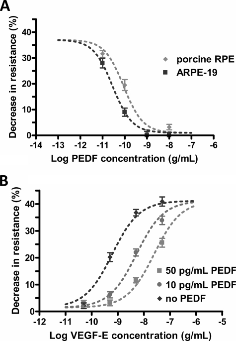
Wet age-related macular degeneration (AMD) attacks the integrity of the retinal pigment epithelium (RPE) barrier system. The pathogenic process was hypothesized to be mediated by vascular endothelial growth factor (VEGF) and antagonized by pigment epithelium-derived factor (PEDF). To dissect these functional interactions, monolayer cultures of RPE cells were established, and changes in transepithelial resistance were evaluated after administration of PEDF, placenta growth factor (VEGF-R1 agonist), and VEGF-E (VEGF-R2 agonist). A recently described mechanism of VEGF inhibition in endothelia required the release of VEGF-R1 intracellular domain by gamma-secretase. To evaluate this pathway in the RPE, cells were pretreated with inhibitors DAPT or LY411575. Processing of VEGF receptors was assessed by Western blot analysis. Administration of VEGF-E rapidly increased RPE permeability, and PEDF inhibited the VEGF-E response dose-dependently. Both gamma-secretase antagonists prevented the inhibitory effects of PEDF. The co-administration of PEDF and VEGF-E depleted the amount of VEGF-R2 in the membrane and increased the amount of VEGF-R2 ectodomain in the media. Therefore, the inhibitory effect of PEDF appears to be mediated via the processing of VEGF-R2 by gamma-secretase. gamma-Secretase generates the amyloid-beta (Abeta) peptide of Alzheimer disease from its precursor (amyloid precursor protein). This peptide is also a component of drusen in dry AMD. The results support the hypothesis that misregulation of gamma-secretase may not only lead to Abeta deposits in dry AMD but can also be damaging to RPE function by blocking the protective effects of PEDF to prevent VEGF from driving the dry to wet AMD transition.


Figures

FIGURE 1.
FIGURE 1.
PEDF inhibits the RPE response to VEGF-E. The RPE response and the inhibition are polarized and VEGF-R2-dependent. Transepithelial resistance measurements on ARPE-19 (A) and porcine primary RPE cell monolayers (B) grown on permeable membrane filters. The cells were treated with apical VEGF-E (5 ng/ml) and apical or basal PEDF (5 ng/ml). The drop in resistance induced by VEGF-E was blocked by only apically administered PEDF. Values are means ± S.E. of individual measurements normalized to the average TER at t = 0 (*, p < 0.01). C, inulin flux through ARPE-19 cell monolayers. The apical to basolateral movement of carboxyl-[14C]inulin was assessed after treatment with apical VEGF (10 ng/ml); apical VEGF (10 ng/ml) and apical PEDF (5 ng/ml); or vehicle. The inulin flux, determined from the slope of the regression analysis, was 570 μg/h for VEGF-PEDF co-treatment, which compares to the 580 μg/h measured for vehicle-treated controls. Because the inulin flux was 780 μg/h after VEGF treatment, these results mean that PEDF prevented the 36% reduction in barrier function 5 h after VEGF administration.
FIGURE 2.
FIGURE 2.
Immunohistochemical localization of VEGF-R2 in sections of porcine eyecups. A–C, images are overlays of simultaneous VEGF-R2 (red, rhodamine-conjugated secondary antibody), nuclear stain (blue), and transmitted-light microscopy photos. Only the apical surface of the RPE is stained for VEGF-R2, but VEGF-R2 staining is abundant in all layers of the neural retina, as seen in A. B, enlargement of the RPE and photoreceptors, to show VEGF-R2 localization in the RPE. C, negative staining control without the anti-VEGF-R2 primary antibody.
FIGURE 3.
FIGURE 3.
Concentration-dependent PEDF response. A, the VEGF-E-induced RPE barrier breakdown depends on the concentration of apical PEDF. ARPE-19 and primary porcine RPE cell monolayers were simultaneously treated with VEGF-E (5 ng/ml) and various concentrations of PEDF (10 pg/ml to 10 ng/ml). The figure shows the percent resistance drop at 2 h relative to the resistance before the treatments. Log IC50 = −10.54 ± 0.06 (29 pg/ml) for ARPE-19 cells and −10.05 ± 0.1 (89 pg/ml) for primary porcine RPE cell monolayers. The Hill coefficients were not significantly different from 1.0. B, Gaddum/Schild competitive model for the interaction of VEGF and PEDF in ARPE-19 cells. Monolayers were treated with various concentrations of VEGF-E (50 pg/ml to 50 ng/ml) and PEDF (0–50 pg/ml). The figure shows the percent resistance drop at 2 h relative to the resistance before the treatments. The data were analyzed with the Gaddum/Schild competitive inhibition model of the entire family of curves resulting in pA2 = 11.85 ± 0.30 and SchildSlope = 1.03 ± 0.22. Thus, PEDF is a competitive inhibitor of VEGF-R2 with Kb = 1.4 ± 0.7 pg/ml in ARPE-19 cell monolayers. Values are the mean ± S.E. normalized to the average resistance at t = 0.
FIGURE 4.
FIGURE 4.
γ-Secretase mediates the PEDF-induced reversal of the RPE response to VEGF. ARPE-19 (A) and primary porcine RPE (B) cell monolayers were pretreated with DAPT (1 μm) or vehicle (0.1% DMSO) 1 h prior to simultaneous PEDF (1 ng/ml) and VEGF-E (5 ng/ml) treatments. VEGF-E (5 ng/ml), PEDF (1 ng/ml), and DAPT (1 μm) were used as controls. The γ-secretase inhibitor, DAPT, prevented PEDF from blocking the VEGF-E-induced TER drop. The controls did not significantly change the resistance. Values are the mean ± S.E. of individual measurements normalized to the TER at t = −60 min (*, p < 0.01). C, the reversal of the effect of PEDF is concentration-dependent. γ-Secretase antagonists, DAPT and LY411575, were utilized in various concentrations (100 pm to 10 μm) to reverse the inhibitory effects of PEDF on RPE barrier breakdown. The figure shows the percent resistance drop at 2 h relative to the resistance before the treatments. Log EC50 was −8.41 ± 0.12 (EC50 = 4 nm) for LY411575 and −7.54 ± 0.12 (IC50 = 29 nm) for DAPT, as LY411575 is a more potent inhibitor than DAPT. The Hill coefficients calculated from the concentration response curves were 0.97 for LY411575 and 0.78 for DAPT.
FIGURE 5.
FIGURE 5.
Co-administration of VEGF and PEDF induces γ-secretase activity. A, γ-secretase activity was probed with a C-terminal APP antibody against the membrane fraction from confluent ARPE-19 cell monolayers. The cells were pretreated with DAPT (1 μM) or vehicle (0.1% DMSO) 1 h prior to PEDF (1 ng/ml) or VEGF-E (5 ng/ml) treatment for 2 h. The lanes represent individual cultures treated by 1, 0.1% DMSO (vehicle); 2, VEGF-E (5 ng/ml); 3, PEDF (1 ng/ml) and VEGF-E (5 ng/ml) and DAPT (1 μm); and 4, PEDF (1 ng/ml) and VEGF-E (5 ng/ml). The gel micrograph shows bands for the full-length APP protein (78 kDa), and the CTFα fragment (11 kDa). B, summary of the γ-secretase activity data. Six independent experiments were performed similar to panel A, and the densities for the APP and CTFα bands were analyzed. The activity of γ-secretase is inversely proportional to the amount of CTFα present. DAPT antagonized γ-secretase activity (lane 3). The PEDF and VEGF-E treatments alone did not significantly change γ-secretase activity from vehicle-treated controls, however, PEDF and VEGF-E together induced a 24% increase in γ-secretase activity. Values are the mean ± S.E. of individual measurements (*, p < 0.05 compared with VEGF treatment). C, the expression of presenilin-1 (PS1) did not significantly change in response to the above treatments. The gel micrograph shows the bands for full-length presenilin-1 (46 kDa). D, the effects of PEDF are not mediated by the catalytic activity of the PEDF receptor. ARPE-19 cell monolayers were pretreated with 25 μm bromoenol lactone (BEL) 1 h prior to PEDF (1 ng/ml), VEGF-E (5 ng/ml), or simultaneous PEDF (1 ng/ml) and VEGF-E (5 ng/ml) treatments. BEL is an inhibitor of the catalytic phospholipase A2 activity of the PEDF receptor in the RPE. The responses to VEGF and/or PEDF are not significantly different from cultures that were not pretreated with BEL. Values are the mean ± S.E. of individual measurements normalized to the TER at t = −60 min (*, p < 0.01).
FIGURE 6.
FIGURE 6.
VEGF-R2 receptor shedding after co-administration of VEGF and PEDF. VEGF-R2 ectodomain appears in the apical media of RPE cells after simultaneous PEDF and VEGF-E treatments. A, Western blot of apical media from ARPE-19 cell monolayers pretreated with DAPT (1 μm) or vehicle (0.1% DMSO) 1 h prior to PEDF (1 ng/ml) or VEGF-E (5 ng/ml) treatments for 2 h and probed with an N-terminal VEGF-R2 antibody. One band was observed at 160 kDa. The lanes represent individual cultures and were treated by 1, VEGF-E (5 ng/ml); 2, PEDF (1 ng/ml); 3, PEDF (1 ng/ml) and VEGF-E (5 ng/ml) and DAPT (1 μm); and 4, PEDF (1 ng/ml) and VEGF-E (5 ng/ml). B, summary of the VEGF-R2 immunoblots. Three independent experiments were performed similar to panel A, and the density for the band (at 160 kDa) was analyzed. PEDF and VEGF-E together induced an increase in VEGF-R2 bands in the media versus the VEGF-E treatment. Pretreatment with DAPT reversed the combined effect of PEDF and VEGF. The values are the mean ± S.E. of individual measurements (*, p < 0.05 compared with control). C, Western blot of apical media from ARPE-19 cell monolayers with an N-terminal VEGF-R1 antibody. One band was observed at 170 kDa. The lanes represent individual cultures and were treated similar to panel A. There was no significant difference between the four treatments, demonstrating that the ectodomain of the VEGF-R1 receptor is not processed after VEGF-E and PEDF treatments. On the other hand, VEGF-R2 is also processed in the cell membranes. D, the cell membranes of ARPE-19 cells (treated similar to panel A) were collected and probed with a C-terminal VEGF-R2 antibody. One band was observed at 220 kDa for the full-length receptor. E, summary of VEGF-R2 immunoblots from the cell membranes. Three independent experiments were performed similarly to panel A, and the density for the band at 220 kDa was analyzed. PEDF and VEGF-E together induced a significant decrease in the VEGF-R2 band. Pretreatment with DAPT reversed the combined effect of PEDF and VEGF. The values are the mean ± S.E. of individual measurements (*, p < 0.05 compared with control). F, Western blot of ARPE-19 cell membranes probed with a C-terminal VEGF-R1 antibody. One band was observed at 250 kDa for the full-length receptor. VEGF-R1 was unaffected by the treatments, demonstrating that it did not mediate the combined effects of VEGF and PEDF.
FIGURE 7.
FIGURE 7.
The proposed mechanism of VEGF-R2 processing. The combined action of PEDF and VEGF activates a yet unknown α-secretase and the known γ-secretase to shed the VEGF-R2 ectodomain and to rapidly clear the receptor from the RPE cell membrane, which eliminates the VEGF-R2-induced permeability increase. The binding of VEGF-E to the released VEGF-R2 ectodomain is responsible for the high affinity competitive inhibition. DAPT blocks γ-secretase; therefore, VEGF-R2 receptor signaling is maintained against the combined actions of PEDF and VEGF. Moreover, the γ-secretase inhibition has a negative feedback on the ectodomain shedding.

Similar articles

Cited by

References

    1. Tranos P. G., Wickremasinghe S. S., Stangos N. T., Topouzis F., Tsinopoulos I., Pavesio C. E. (2004) Surv. Ophthalmol. 49, 470–490 - PubMed
    1. Augustin A. J., Puls S., Offermann I. (2007) Retina 27, 133–140 - PubMed
    1. Marmor M. F. (1999) Documenta Ophthalmologica 97, 239–249 - PubMed
    1. Gragoudas E. S., Adamis A. P., Cunningham E. T., Jr., Feinsod M., Guyer D. R. (2004) N. Engl. J. Med. 351, 2805–2816 - PubMed
    1. Heier J. S., Antoszyk A. N., Pavan P. R., Leff S. R., Rosenfeld P. J., Ciulla T. A., Dreyer R. F., Gentile R. C., Sy J. P., Hantsbarger G., Shams N. (2006) Ophthalmology 113, 642, e641–644 - PubMed

Publication types

MeSH terms

Substances