. 2010 Jan;24(1):275-85.
doi: 10.1096/fj.09-137158. Epub 2009 Sep 1.

Germline deletion of AMP-activated protein kinase beta subunits reduces bone mass without altering osteoclast differentiation or function

Affiliations

Germline deletion of AMP-activated protein kinase beta subunits reduces bone mass without altering osteoclast differentiation or function

Julian M W Quinn et al. FASEB J. 2010 Jan.

Abstract

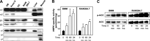
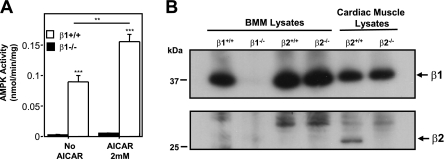
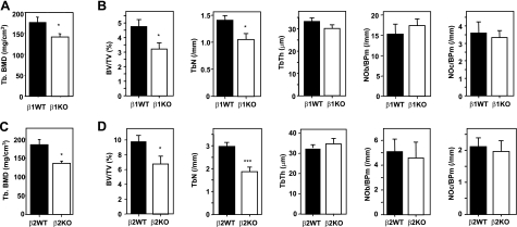
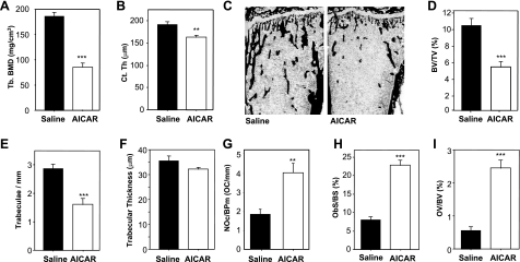
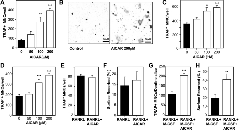
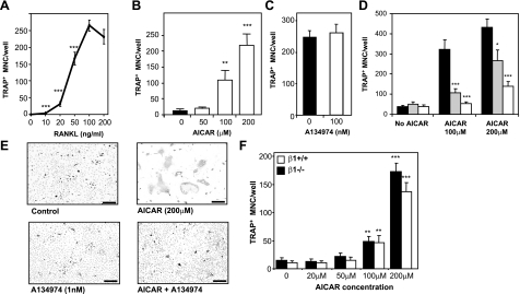
Since AMP-activated protein kinase (AMPK) plays important roles in modulating metabolism in response to diet and exercise, both of which influence bone mass, we examined the influence of AMPK on bone mass in mice. AMPK is an alphabetagamma heterotrimer where the beta subunit anchors the alpha catalytic and gamma regulatory subunits. Germline deletion of either AMPK beta1 or beta2 subunit isoforms resulted in reduced trabecular bone density and mass, but without effects on osteoclast (OC) or osteoblast (OB) numbers, as compared to wild-type littermate controls. We tested whether activating AMPK in vivo would enhance bone density but found AICA-riboside treatment caused a profound loss of trabecular bone volume (49.5%) and density and associated increased OC numbers. Consistent with this, AICA-riboside strongly stimulated OC differentiation in vitro, in an adenosine kinase-dependent manner. OCs and macrophages (unlike OBs) lacked AMPK beta2 subunit expression, and when generated from AMPK beta1(-/-) mice displayed no detectable AMPK activity. Nevertheless, AICA-riboside was equally effective at stimulating OC differentiation from wild-type or beta1(-/-) progenitors, indicating that AMPK is not essential for OC differentiation or the stimulatory action of AICA-riboside. These results show that AMPK is required to maintain normal bone density, but not through bone cell differentiation, and does not mediate powerful osteolytic effects of AICA-riboside.


Figures

Figure 1.
Figure 1.
AMPK subunit expression and activation in bone cells. A) Western blot analysis of subunit expression. AMPK heterotrimers were immunoabsorbed from cell lysates using α1/α2 specific antibodies. Blots were probed with AMPK subunit isoform-specific antibodies listed at left. Cell lysates included primary calvarial OBs, Kusa O cells, in vitro generated OC-containing cultures, and RAW 264.7 cells. Mouse liver and skeletal muscle (or cardiac for γ2) lysates were included as controls. B) AICAR activation of AMPK in BMMs and RAW264.7 cells. Cells were stimulated with 2 mM AICAR for 10 and 60 min, and 0.2 mM for 24 h time points. AMPK heterotrimers were immunoprecipitated from cell lysates, and AMPK activity was measured by SAMS peptide phosphorylation assay. C) Phosphorylation of AMPK target ACC resulting from AICAR treatment. ACC was precipitated by biotin affinity from cell lysates, and ACC total and phosphorylated ACC (p-ACC) was detected by Western blotting methods. *P < 0.05 vs. positive controls (black columns).
Figure 2.
Figure 2.
BMMs derived from AMPK β1−/− mice are AMPK null. A) Lack of AMPK activity in BMMs derived from AMPK β1−/− mice. BMMs were treated with or without AICAR (2 mM, 30 min) to activate AMPK. AMPK activity was measured as outlined in Materials and Methods. **P < 0.01, ***P < 0.001 vs. respective negative control. B) AMPKβ1−/− BMMs do not express a β2 subunit. BMMs and cardiac muscle tissue lysates prepared from AMPK β1 and β2 wild-type (+/+) and knockout mice (−/−) were immunoabsorbed using α1/α2 antibodies. Western blots were probed with a monoclonal antibody recognizing both β1 and β2 subunits (Epitomics). β1 is expressed in all BMM lysates (except β1−/− BMMs) as well as cardiac muscle. β2 is expressed in cardiac lysates from β2+/+ mice but not β2−/− mice and is absent from all BMM lysates. See Supplemental Material for details describing the generation and preliminary characterization of AMPKβ2−/− null mice.
Figure 3.
Figure 3.
Bone phenotypes of germline AMPK β1- and β2-subunit-knockout mice. A) pQCT analysis of trabecular bone of femora of female WT and AMPKβ1−/− mice. B) Histomorphometric analysis of female AMPKβ1−/− mouse bones. C) pQCT analysis of trabecular bone of femora of male WT and AMPKβ2−/− mice. D) Histomorphometric analysis of male WT and AMPKβ2−/− mouse bones. *P < 0.05, ***P < 0.001 vs. WT (black columns).
Figure 4.
Figure 4.
AICAR causes bone loss and elevated bone turnover. Male C57Bl/6 mice (10 wk old) were treated with AICAR (500 mg/kg/d) or saline for 4 wk. A, B) pQCT analysis of femoral trabecular density (A) and femoral cortical thickness (B). C) Low-power views of undecalcified sections of tibia from saline and AICAR-treated mice histochemically stained by von Kossa method (black indicates mineralized bone) and counterstained with ponceau-orange G. D) Tibial trabecular volume, BV/TV. E) Tibial trabecular numbers/mm (Tb.N). F) Tibial trabecular thickness (Tb.Th). G) Tibial OC numbers on trabecular bone (Oc.N). H) Surface of tibial bone occupied by OBs, Ob.S/BS. I) Tibial osteoid volume (OV/BV). **P < 0.01, ***P < 0.001 vs. saline control (black columns); n = 6 mice/treatment group.
Figure 5.
Figure 5.
AICAR causes supramaximal OC formation in vitro without effects on OC survival or activity. A) Dose-response effect of AICAR on OC formation in cocultures of bone marrow cells with calvarial OBs, stimulated by 10–8 M 1,25(OH)2-D3 for 7 d. B) Photomicrographs of cocultures after staining for TRAP (dark gray in image), showing control and 200 μM AICAR-treated cells. C) Dose-response effect of AICAR on OC formation in bone marrow cells maximally stimulated by 100 ng/ml RANKL and 30 ng/ml M-CSF. D) Dose response of AICAR on OC formation in 100 ng/ml RANKL-stimulated RAW264.7 cells. E) AICAR (200μM) effects on 24 h survival of RANKL-treated dispersed OCs. F) AICAR (200 μM) effects on dentine resorption area by 100 ng/ml RANKL-stimulated dispersed OCs incubated for 3 d. G) Numbers of OCs formed on dentine in cultures of bone marrow cells stimulated for 10 d by RANKL (100 ng/ml) and M-CSF (30 ng/ml) in the presence and absence of AICAR. H) AICAR effects on resorption by OCs formed on dentine, as in G. **P < 0.01, ***P < 0.001 vs. positive control (black columns).
Figure 6.
Figure 6.
AICAR-increased OC formation is ZMP dependent but AMPK independent. A) Dose response of RANKL on OC formation. ***P < 0.01 vs. 100 ng/ml RANKL. B) Dose response of AICAR on OC formation in bone marrow cells stimulated with submaximal RANKL (20 ng/ml) and 30 ng/ml M-CSF. C) Effects of A134974 on bone marrow cell cultures maximally stimulated by RANKL (100 ng/ml) and M-CSF (30 ng/ml). D) Ablation of AICAR action on OC formation by A134974; bone marrow cell cultures submaximally stimulated with RANKL (20 ng/ml) and 30 ng/ml M-CSF in presence and absence of AICAR. Gray columns, 100 pM A134974; white columns, 1 nM A134974. E) Photomicrographs of TRAP-positive cells (dark gray) in bone marrow cell cultures stimulated with submaximal (20 ng/ml) RANKL, as in B; control, A134974 treated, AICAR-treated, and AICAR plus A134974-treated cultures. Scale bars = 50 μm. F) Effects of AICAR treatment on OC formation from 20 ng/ml RANKL-stimulated BMMs from AMPKβ1−/− and AMPKβ1+/+ mice. *P < 0.05, **P < 0.01, ***P < 0.001 vs. respective controls.

References

    1. Karsenty G. Convergence between bone and energy homeostases: leptin regulation of bone mass. Cell Metab. 2006;4:341–348. - PubMed
    1. Seibel M J. Nutrition and molecular markers of bone remodelling. Curr Opin Clin Nutr Metab Care. 2002;5:525–531. - PubMed
    1. Scott J W, Oakhill J S, van Denderen B J W. AMPK/SNF1 structure: a ménage a trois of energy-sensing. Front Biosci. 2009;14:596–610. - PubMed
    1. Sun W, Lee T S, Zhu M, Gu C, Wang Y, Zhu Y, Shyy J Y. Statins activate AMP-activated protein kinase in vitro and in vivo. Circulation. 2006;114:2655–2662. - PubMed
    1. Gutierrez G E, Edwards J R, Garrett I R, Nyman J S, McCluskey B, Rossini G, Flores A, Neidre D B, Mundy G R. Transdermal lovastatin enhances fracture repair in rats. J Bone Miner Res. 2008;23:1722–1730. - PMC - PubMed

Publication types

MeSH terms