. 2010 Jan;24(1):296-308.
doi: 10.1096/fj.09-135087. Epub 2009 Sep 1.

Antiapoptotic roles of ceramide-synthase-6-generated C16-ceramide via selective regulation of the ATF6/CHOP arm of ER-stress-response pathways

Affiliations

Antiapoptotic roles of ceramide-synthase-6-generated C16-ceramide via selective regulation of the ATF6/CHOP arm of ER-stress-response pathways

Can E Senkal et al. FASEB J. 2010 Jan.

Abstract

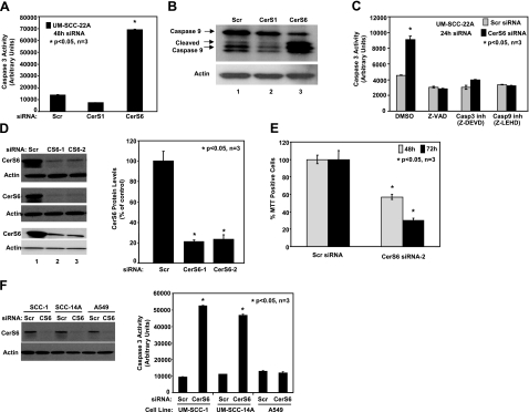
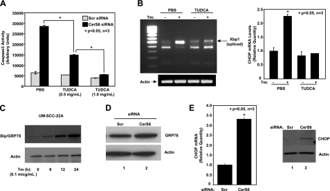
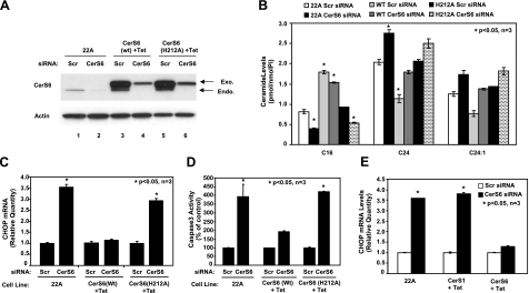
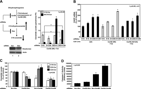
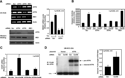
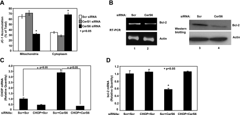
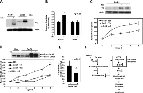
Emerging results suggest that ceramides with different fatty acid chain lengths might play distinct functions in the regulation of tumor growth and therapy. Here we report that de novo-generated C(18)- and C(16)-ceramides by ceramide synthases 1 and 6 (CerS1 and CerS6) play opposing proapoptotic and prosurvival roles, respectively, in human head and neck squamous cell carcinomas (HNSCCs). Unexpectedly, knockdown of CerS6/C(16)-ceramide using small interfering RNA induced endoplasmic reticulum (ER)-stress-mediated apoptosis. Reconstitution of C(16)-ceramide generation by induced expression of wild-type CerS6, but not its catalytically inactive mutant, protected cells from cell death induced by knockdown of CerS6. Moreover, using molecular tools coupled with analysis of sphingolipid metabolism showed that generation of C(16)-ceramide, and not dihydro-C(16)-ceramide, by induced expression of CerS6 rescued cells from ER stress and apoptosis. Mechanistically, regulation of ER-stress-induced apoptosis by CerS6/C(16)-ceramide was linked to the activation of a specific arm, ATF6/CHOP, of the unfolded protein response pathway. Notably, while expression of CerS1/C(18)-ceramide inhibited HNSCC xenograft growth, CerS6/C(16)-ceramide significantly protected ER stress, leading to enhanced tumor development and growth in vivo, consistent with their pro- and antiapoptotic roles, respectively. Thus, these data reveal an unexpected and novel prosurvival role of CerS6/C(16)-ceramide involved in the protection against ER-stress-induced apoptosis and induction of HNSCC tumor growth.


Figures

Figure 1.
Figure 1.
Knockdown of CerS6 induces caspase activation and cell death. A, B) UM-SCC-22A cells were transfected with either scrambled (Scr), CerS1, or CerS6 siRNAs at 50 nM for 48 h, and their effects on the activation of caspase 3 (A) or caspase 9 (B) were measured. C) Roles of caspase inhibitors Z-VAD, Z-DEVD, and Z-LEHD in the prevention of caspase 3 activation in response to knockdown of CerS6 were examined. D) Left panel: down-regulation of CerS6 using siRNAs was confirmed by Western blotting in 3 independent experiments. Right panel: CerS6 protein levels were quantified from the blots and normalized to the levels of actin. E) Effects of knockdown of CerS6 using CerS6 siRNA-2 on UM-SCC-22A cell growth were measured by MTT after 48 and 72 h of transfection. F) UM-SCC-1, UM-SCC-14A, and A549 cells were transfected with either Scr or CerS6 siRNA (50 nM, 48 h); down-regulation of CerS6 was confirmed by Western blotting (F, left panel); and caspase 3 activity was measured (F, right panel). Data represent 3 independent studies. Bars represent means ± sd from 3 independent experiments. *P < 0.05.
Figure 2.
Figure 2.
Down-regulation of CerS6 induces ER-stress-mediated apoptosis. A) UM-SCC-22A cells were transfected with either Scr or CerS6 siRNA (50 nM, 48 h) in the presence of either vehicle (PBS) or TUDCA (0.5 and 1 mg/ml), and caspase 3 activity was measured. Bars represent means ± sd from 3 independent experiments. *P < 0.05. B) UM-SCC-22A cells were pretreated with TUDCA (1 mg/ml, 2 h) and treated with tunicamycin (Tm; 0.1 μg/ml, 2 h). After treatments, XBP-1 mRNA splicing was analyzed using RT-PCR (left panel); CHOP mRNA levels were measured by Q-RT-PCR (right panel). C) UM-SCC-22A cells were treated with tunicamycin at 0.1 μg/ml for indicated time points, and its effects on Bip/Grp78 and actin protein levels were examined by Western blotting. D, E) UM-SCC-22A cells were transfected with either Scr or CerS6 siRNA (50 nM, 48 h), and protein levels of Bip/GRP78 were detected by Western blotting (D), or mRNA and protein levels of CHOP were measured using Q-RT-PCR (E, left panel) and Western blotting (E, right panel), respectively.
Figure 3.
Figure 3.
Prevention of CHOP mRNA induction by reconstitution of C16-dh-ceramide generation via expression of wt-CerS6. AD) Control UM-SCC-22A (22A) cells (lanes 1, 2), and Tet-induced (+Tet) UM-SCC-22A cells expressing either WT (lanes 3, 4) or catalytically inactive mutant (H212A) CerS6 (lanes 5, 6) were transfected with either Scr or CerS6 siRNA (50 nM) for 48 h, and expression levels of CerS6 or actin proteins (A) were detected by Western blotting. Then, their effects on ceramide levels were measured by LC/MS (B), CHOP mRNA was measured by Q-PCR (C), and activation of caspase 3 was measured by activity assay (D). E) Tet-induced cells expressing CerS1 or CerS6 were transfected with siRNA as in A, and CHOP mRNA was measured by Q-PCR. Bars represent means ± sd from 3 experiments. *P < 0.05.
Figure 4.
Figure 4.
Dissection of the roles of dihydroceramide or ceramide in the regulation of ER stress. A) Left panel: schematic representation of the experiments carried out to dissect the roles of C16- or dh-C16-ceramides in the prevention of CHOP activation and ER stress. Right panel: C16- and dh-C16-ceramide levels of Tet-induced UM-SCC-22A cells expressing CerS6 (WT) after transfections with Scr, Des, or CerS6 siRNAs were measured by LC/MS. Down-regulation of Des using siRNA in UMSCC-22A cells compared with nontargeting scrambled siRNA-treated cells was confirmed by Western blotting (bottom panel, lanes 2 and 1, respectively). Actin levels were used as controls. B) Control UM-SCC-22A (22A) cells and Tet-induced (+Tet) UM-SCC-22A cells expressing either WT or catalytically inactive mutant (H212A) CerS6 were transfected with Scr (S), Des (D), or CerS6 (CS6) siRNAs (20 nM) alone or in combination (S+S, D+S, S+CS6, and D+CS6) for 48 h, and their roles in the regulation of CHOP mRNA levels were measured by Q-PCR. C, D) Roles of down-regulation of CerS2 and CerS6, alone or in combination, in the regulation of ceramide levels (C) and activation of caspase 3 (D) were detected by LC/MS and caspase activity assay, respectively. Bars represent means ± sd from 2 experiments done in duplicates. *P < 0.05.
Figure 5.
Figure 5.
Down-regulation of CerS6 specifically activates the ATF6/CHOP arm of ER-stress response pathways. A) Left panels: specific knockdown of ATF4, ATF6, and IRE1 using siRNAs was confirmed using RT-PCR (top panel) and by Western blotting (bottom panel). Right panel: functional down-regulation of ATF4 in response to siRNA transfection was confirmed by analysis of REDD1 mRNA levels by Q-RT-PCR in the absence and presence of tunicamycin (Tm). B, C) Roles of ATF4, ATF6, and IRE1 in the regulation of caspase 3 activation (B) or CHOP mRNA (C) in response to down-regulation of CerS6 were examined. D) Roles of down-regulation of CerS6 in the regulation of ATF6 activation in UM-SCC-22A cells were examined, following transfections with N-terminal FLAG-tagged ATF6 and/or CerS6 siRNA. Cleaved and uncleaved ATF6 were detected by anti-FLAG immunoprecipitation followed by immunoblotting (left panel). Data represent 3 independent experiments. Band intensities were quantified and plotted as cleaved to full-length ATF6 ratio (right panel). Bars represent means ± sd from at least 2 experiments. *P < 0.05.
Figure 6.
Figure 6.
Knockdown of CerS6 down-regulates Bcl-2 via activation of CHOP and induces loss of mitochondrial membrane potential. A) UM-SCC-22A cells were transfected with Scr, CerS1, or CerS6 siRNA (50 nM, 48 h), and their effects on the loss of mitochondrial membrane potential were detected by JC-1 staining followed by flow cytometry. B) UM-SCC-22A cells were transfected with Scr or CerS6 siRNA (50 nM, 48 h), and their effects on the mRNA and protein levels of Bcl-2 were examined by RT-PCR and Western blotting. C, D) Roles of CerS6 and CHOP in the down-regulation of Bcl-2 mRNA by RT-PCR were examined in UM-SCC-22A cells, after knockdown of CerS6 and/or CHOP, alone or in combination using siRNAs. CHOP (C) and Bcl-2 (D) mRNA levels were measured by Q-RT-PCR. Results represent 2 independent experiments performed in duplicate. *P < 0.05.
Figure 7.
Figure 7.
Roles of CerS1 and CerS6 in the regulation of HNSCC tumor growth. A) UM-SCC-22A cells expressing C-terminal V5-tagged CerS1 or CerS6 under a Tet-inducible promoter were either noninduced (−Tet) or induced with Tet (1 μg/ml; +Tet) for 48 h, and CerS1 and CerS6 protein expression was detected by Western blotting using V5 antibody. Untransfected UM-SCC-22A cells (22A) were used as controls. B) Activation of caspase 3 was measured without or with Tet induction (48 h) in cells expressing CerS1 or CerS6. Bars represent means ± sd from 3 experiments. C–E) HNSCC xenografts were generated on the flanks of SCID mice using control UM-SCC-22A (22A) cells and UM-SCC-22A cells expressing CerS1 or CerS6 under a Tet-inducible promoter, as described in Materials and Methods. Sizes of the xenografts were measured every 3 d with calipers. Mice with the described xenografts were sacrificed, and CerS1 and actin protein (C), CerS6 (D), and CHOP (E) levels of the xenografts were detected by Western blotting and by Q-PCR. Bars represent means ± sd. *P < 0.05. F) Summary of data presented in this study. Results revealed that knockdown of CerS6 using siRNA initiates a specific arm of the ER stress response through the ATF6/CHOP pathway, which then leads to induction of caspase-dependent apoptosis.

References

    1. Ogretmen B, Hannun Y A. Biologically active sphingolipids in cancer pathogenesis and treatment. Nat Rev Cancer. 2004;4:604–616. - PubMed
    1. Mehta S, Blackinton D, Omar I, Kouttab N, Myrick D, Klostergaard J, Wanebo H. Combined cytotoxic action of paclitaxel and ceramide against the human Tu138 head and neck squamous carcinoma cell line. Cancer Chemother Pharmacol. 2000;46:85–92. - PubMed
    1. Senkal C E, Ponnusamy S, Rossi M J, Bialewski J, Sinha D, Jiang J C, Jazwinski S M, Hannun Y A, Ogretmen B. Role of human longevity assurance gene 1 and C18-ceramide in chemotherapy-induced cell death in human head and neck squamous cell carcinomas. Mol Cancer Ther. 2007;6:712–722. - PubMed
    1. Swanton C, Marani M, Pardo O, Warne P H, Kelly G, Sahai E, Elustondo F, Chang J, Temple J, Ahmed A A, Brenton J D, Downward J, Nicke B. Regulators of mitotic arrest and ceramide metabolism are determinants of sensitivity to paclitaxel and other chemotherapeutic drugs. Cancer Cell. 2007;11:498–512. - PubMed
    1. Guillas I, Kirchman P A, Chuard R, Pfefferli M, Jiang J C, Jazwinski S M, Conzelmann A. C26-CoA-dependent ceramide synthesis of Saccharomyces cerevisiae is operated by Lag1p and Lac1p. EMBO J. 2001;20:2655–2665. - PMC - PubMed

Publication types

MeSH terms