Review
. 2009 Sep 1;23(17):1986-97.
doi: 10.1101/gad.1819909.

Signals and prepatterns: new insights into organ polarity in plants

Affiliations
Review

Signals and prepatterns: new insights into organ polarity in plants

Aman Y Husbands et al. Genes Dev. .

Abstract

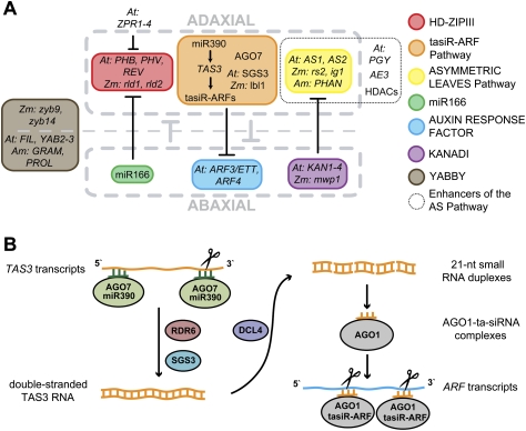
The flattening of leaves results from the interaction between upper (adaxial) and lower (abaxial) domains in the developing primordium. These domains are specified by conserved, overlapping genetic pathways involving several distinct transcription factor families and small regulatory RNAs. Polarity determinants employ a series of antagonistic interactions to produce mutually exclusive cell fates whose positioning is likely refined by signaling across the adaxial-abaxial boundary. Signaling candidates include a mobile small RNA-the first positional signal described in adaxial-abaxial polarity. Possible mechanisms to polarize the incipient primordium are discussed, including meristem-derived signaling and a model in which a polarized organogenic zone prepatterns the adaxial-abaxial axis.


Figures

Figure 1.
Figure 1.
Adaxial–abaxial leaf polarity. (A) The adaxial side of an Arabidopsis leaf is dark green and trichome-rich, whereas the abaxial leaf surface is gray-green and trichome-poor. (B) Transverse section through a Nerium leaf illustrating the differentiation of distinct cell types within the adaxial and abaxial domains. Rectangular palisade mesophyll (P) cells form a tightly packed file beneath the adaxial epidermis, whereas spongy mesophyll (S) cells separated by large intercellular air spaces differentiate abaxially. Within the vasculature (V), water-bearing xylem cells differentiate adaxial to sugar-bearing phloem cells. (C) Longitudinal section of an Arabidopsis apex, showing the proximal–distal and adaxial–abaxial axes of leaf primordia relative to the SAM (marked M). (D) Transverse section of an Arabidopsis vegetative shoot apex, showing the positions of the adaxial and abaxial sides of leaf primordia relative to the meristem (M). Note the spiral phyllotaxis of leaves around the SAM with increasingly older primordia at a greater distance from the meristem. (The image in B is used with permission from the University of Wisconson Plant Teaching Collection at http://botit.botany.wisc.edu/images/130/Leaf.)
Figure 2.
Figure 2.
Phenotypes of mutants with perturbed adaxial–abaxial patterning. (A) Normal maize leaves develop flattened blades due to interaction between the adaxial and abaxial domains. lbl1 mutants interfere with ta-siRNA biogenesis and adaxial cell fate specification and consequently their leaves are often radial and abaxialized. (B) Transverse sections through a wild-type (top), weakly adaxialized mwp1 (middle), and fully abaxialized lbl1 (bottom) leaf. Note the formation of ectopic blade outgrowths at the boundaries of adaxialized tissue sectors on the abaxial leaf surface of the mwp1 leaf (marked by orange lines), and the radial symmetry of the lbl1 leaf. (Orange lines) Adaxial; (blue lines) abaxial. (The images in B are reproduced with permission from Candela et al. [2008] [© 2008 American Society of Plant Biologists] and Timmermans et al. [1998].)
Figure 3.
Figure 3.
A network of conserved transcription factors and small RNA pathways maintains adaxial–abaxial polarity. (A) The HD-ZIPIII, AS, and TAS3 ta-siRNA pathways contribute to the specification of adaxial cell fate. In Arabidopsis, HD-ZIPIII activity is regulated via a negative feedback loop involving the ZPR proteins, while the PIGGYBACK ribosomal proteins (PGY) (Pinon et al. 2008), ASYMMETRIC LEAVES ENHANCER3 (AE3) (Huang et al. 2006), and histone deacetylase proteins (HDAC) (Ueno et al. 2007) enhance the AS pathway (dotted outline). Members of the KANADI and ARF families, together with the miRNA miR166, contribute to the specification of abaxial identity. The site of YABBY activity varies between species but its contribution to organ outgrowth may be conserved. Antagonistic interactions between the polarity determinants create mutually exclusive adaxial and abaxial cell fates that contribute to the stable maintenance of organ polarity throughout development. Direct interactions are marked with a bold line. (B) Diagram of the TAS3 ta-siRNA pathway. miR390-loaded AGO7 targets TAS3 transcripts, which upon cleavage of the 3′ target site are converted into dsRNAs through the activities of RDR6 and SGS3/LBL1 and subsequently processed by DCL4 into phased 21-nt species. The TAS3-derived ta-siRNAs, tasiR-ARFs, act in trans to repress the expression of the abaxial determinants ARF3 and ARF4.
Figure 4.
Figure 4.
Diverse contributions of TAS3 ta-siRNAs to adaxial–abaxial patterning. (A, top graph) In Arabidopsis, mature miR390 (dark green) accumulates throughout the leaf, but its activity (gray-green box) is restricted to the adaxial side by localized expression of AGO7 (light green). (Middle graph) tasiR-ARF biogenesis (pale orange box) is further confined to the two most adaxial cell layers by the restricted expression of the TAS3A precursor (brown). Mobility of tasiR-ARFs (orange) creates a gradient of accumulation across the developing leaf that is strongest near its adaxial site of biogenesis. (Bottom graph) This gradient yields regions of high and low tasiR-ARF activity, perhaps patterned in part via adaxial expression of PHN/ZLL (gray box), that restrict ARF3 protein accumulation (dark blue) to the abaxial side even though ARF3 transcripts (light blue) are present throughout leaf primordia. (B) The mutually opposing nature of adaxial and abaxial cell fates, resulting in part from antagonistic interactions between cell-autonomous polarity determinants, contributes to the maintenance of organ polarity, but is unlikely sufficient to define a precise boundary between adaxial and abaxial organ domains. Superimposed intercellular signals that act between the domains can provide positional inputs to refine the adaxial–abaxial boundary. The adaxially derived mobile tasiR-ARFs are candidates for such a signal and may act in conjunction with positional signals from the abaxial domain. (C) In maize, miR390 and tasiR-ARFs (orange) accumulate adaxially in initiating and developing leaf primordia. In Arabidopsis, tasiR-ARFs move from their site of biogenesis below the SAM into the meristem proper and therefore accumulate uniformly throughout incipient primordia (IL).
Figure 5.
Figure 5.
Model for adaxial–abaxial axis specification in the incipient primordium. The SAM comprises a stem cell-containing CZ and organogenic PZ. The PZ is envisioned to be patterned into apical/centric (gray yellow) and basal/outer (gray blue) regions based on positional information inherent to the SAM, possibly signals derived from the CZ and cells basal to the PZ. A lateral organ initiates at the site where an auxin maximum (green circles) overlaps the boundary between these PZ regions (I1). The same boundary also prepatterns the incipient organ into adaxial and abaxial domains. A meristem-derived adaxializing signal (pink arrows) is proposed to maintain this initial polarity until the P2 stage of organ development when maintenance mechanisms within the organ are in place. Compound-leafed species may similarly initiate leaflets (lft) at sites where auxin maxima overlap an adaxial–abaxial boundary at the margins of developing primordia (black arrowheads).

References

    1. Addo-Quaye C, Eshoo T, Bartel D, Axtell M. Endogenous siRNA and miRNA targets identified by sequencing of the Arabidopsis degradome. Curr Biol. 2008;18:758–762. - PMC - PubMed
    1. Adenot X, Elmayan T, Lauressergues D, Boutet S, Bouche N, Gasciolli V, Vaucheret H. DRB4-dependent TAS3 trans-acting siRNAs control leaf morphology through AGO7. Curr Biol. 2006;16:927–932. - PubMed
    1. Allen E, Xie Z, Gustafson A, Carrington J. microRNA-directed phasing during trans-acting siRNA biogenesis in plants. Cell. 2005;121:207–221. - PubMed
    1. Alvarez J, Pekker I, Goldshmidt A, Blum E, Amsellem Z, Eshed Y. Endogenous and synthetic microRNAs stimulate simultaneous, efficient, and localized regulation of multiple targets in diverse species. Plant Cell. 2006;18:1134–1151. - PMC - PubMed
    1. Barkoulas M, Hay A, Kougioumoutzi E, Tsiantis M. A developmental framework for dissected leaf formation in the Arabidopsis relative Cardamine hirsuta. Nat Genet. 2008;40:1136–1141. - PubMed

Publication types

MeSH terms

LinkOut - more resources