. 2009 Sep 1;23(17):2076-87.
doi: 10.1101/gad.1788109.

PLZF is a regulator of homeostatic and cytokine-induced myeloid development

Affiliations

PLZF is a regulator of homeostatic and cytokine-induced myeloid development

Sergei Doulatov et al. Genes Dev. .

Abstract

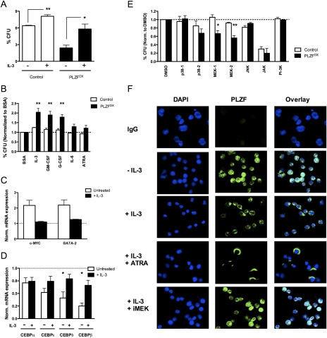
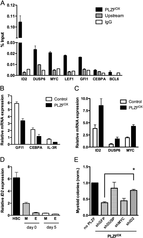
A major question in hematopoiesis is how the system maintains long-term homeostasis whereby the generation of large numbers of differentiated cells is balanced with the requirement for maintenance of progenitor pools, while remaining sufficiently flexible to respond to periods of perturbed cellular output during infection or stress. We focused on the development of the myeloid lineage and present evidence that promyelocytic leukemia zinc finger (PLZF) provides a novel function that is critical for both normal and stress-induced myelopoiesis. During homeostasis, PLZF restricts proliferation and differentiation of human cord blood-derived myeloid progenitors to maintain a balance between the progenitor and mature cell compartments. Analysis of PLZF promoter-binding sites revealed that it represses transcription factors involved in normal myeloid differentiation, including GFI-1, C/EBPalpha, and LEF-1, and induces negative regulators DUSP6 and ID2. Loss of ID2 relieves PLZF-mediated repression of differentiation identifying it as a functional target of PLZF in myelopoiesis. Furthermore, induction of ERK1/2 by myeloid cytokines, reflective of a stress response, leads to nuclear export and inactivation of PLZF, which augments mature cell production. Thus, negative regulators of differentiation can serve to maintain developmental systems in a primed state, so that their inactivation by extrinsic signals can induce proliferation and differentiation to rapidly satisfy increased demand for mature cells.


Figures

Figure 1.
Figure 1.
PLZF expression and viral vector design. (A) Expression of PLZF mRNA in human hematopoietic subsets, before and after culture, was quantified by qPCR. Hematopoietic subsets were isolated by FACS-sorting from fresh Lin CB (day 0) or after 5 d in serum-free culture (day 5) based on the following phenotypes: HSCs, CD34+ CD38, erythroid progenitors (E), CD34+ CD38+ CD71+, myeloid progenitors (M), and CD34+ CD38+ CD71. (B) Schematic representation of viral vectors used to overexpress (MPG, retroviral; CEP, lentiviral) or silence human PLZF (shPLZF). (C) Fold overexpression of PLZF in myeloid progenitors transduced with MPG-PLZF and cultured for 5 d, compared with same-day control cells (M, d5), or freshly sorted myeloid progenitors (M, d0) or HSCs (HSC, d0) from the same CB. (D, left panel) Western blot analysis of total protein extracts from 293T cells transduced with control or MPG-PLZF viruses. (Right panel) Silencing vectors (shPLZF1–3) were transduced into 293T cells stably expressing PLZF. Equal total protein was loaded in all lanes and the blots were probed with an anti-PLZF antibody.
Figure 2.
Figure 2.
PLZF restricts myeloid proliferation and differentiation of human progenitors in vitro. (A) Effect of PLZF on progenitor proliferation. Growth of CD34+ progenitors transduced with MPG (control) or MPG-PLZF (PLZFOX) vectors in serum-free and serum-supplemented cultures quantified by total cell counting. (B) Effect of PLZF on apoptosis. Proportion of annexin V+ (7AAD) apoptotic cells after 7 d in serum-supplemented cultures initiated with control or PLZFOX CD34+ cells; the difference between groups is not significant. (C) Effect of PLZF on cell cycle kinetics. BrdU incorporation of control and PLZFOX CD34+ cells cultured for 7 d in serum-free conditions; representative flow cytometric profiles (left panel) and the proportion of cells in G0/G1, S, and G2/M phases of the cell cycle (right panel) are shown. (D) Effect of PLZF on myeloid differentiation. Proportion of nonmyeloid (CD15 CD14 CD11b) cells in serum-free cultures initiated with control or PLZFOX CD34+ cells. (E) Production of mature lineage-positive myeloid cells in serum-free cultures initiated with sorted control or PLZFOX CD34+ cells. (F) Effect of PLZF on clonogenic progenitors. Colony-forming capacity (colonies counted as proportion of input; percentage of CFU) of freshly sorted control or PLZFOX CD34+ cells. (BFU-E) Erythroid blast-forming units; (CFU-G/M) granulocyte or monocyte colony-forming units; (CFU cells) total myeloid cells × 105 in a CFU-G/M assay. All data are expressed as mean ± SEM of three independent experiments (CB samples), except F, which has four experiments. (*) P < 0.05; (**) P < 0.005.
Figure 3.
Figure 3.
PLZF negatively regulates myeloid development in vivo as assessed in the NOD/SCID xenotransplant system. (A) Effect of PLZF on CD45+ human cell engraftment. Proportion of GFP+ BMCs in individual mice each denoted with a symbol at 8 wk post-transplant (mean marked as solid lines) compared with mean GFP positivity of input HSCs prior to transplantation (broken lines). Lin CB cells were transduced with control (clear triangles), PLZF knockdown (PLZFKD), or CEP-PLZF (PLZFOX) vectors (black triangles) and transplanted into NOD/SCID mice. The level of transduction in three independent experiments ranged from 5% to 12%; as a result, mice were transplanted with a mixture of transduced and nontransduced cells. Thus, equivalent means in the values of the pretransplant and post-transplant percentage of GFP in each experiment indicates a lack of competitive advantage of GFP+ cells relative to nontransduced cells, while increased or decreased percentage of GFP+ indicates competitive advantage or disadvantage, respectively, in vivo. (B) Frequency of human myeloid BMCs in mice transplanted with PLZFKD cells (right) and a representative flow analysis (left). The frequency of granulocytes was calculated as the proportion of SSChi CD33lo cells within the GFP+ CD45+ graft and monocytes as SSClo CD33hi cells within the GFP+ CD45+ graft; the remaining cells were SSClo CD33 lymphocytes. To obtain enough cells for accurate flow cytometric analysis, bone marrow was pooled from four to eight mice. Data are expressed as mean ± SEM of five independent experiments. (**) P < 0.01. (C) Expansion of the absolute number of human granulocytes, monocytes, and lymphoid cells in mice transplanted with PLZFKD cells normalized to controls. The absolute number was calculated by multiplying the frequency of each cell type by the number of GFP+ CD45+ cells in both femurs, tibiae, and pelvis. Data are expressed as mean ± SEM of five independent experiments. (*) P < 0.02. (D) Representative analysis of the proportion of CD11b+ CD16+ human neutrophils in the GFP+ fraction of mice engrafted with Lin CB cells transduced with control, PLZFKD, or PLZFOX viruses. To obtain enough cells for accurate flow cytometric analysis, bone marrow was pooled from four to eight mice.
Figure 4.
Figure 4.
PLZF expands the human progenitor compartment in vivo. (A) Proportion of GFP+ cells within the total human CD45+ graft in the bone marrow (left) compared with Lin CD34+ progenitor fraction (right) in a representative experiment 8 wk post-transplant. Bone marrow was pooled from four to eight mice, depleted of murine and mature human cells, and analyzed for GFP and CD34 expression by flow cytometry before and after lineage depletion. Equal percentage of GFP indicates equal contribution of transduced cells to mature and progenitor compartments, while increased or decreased proportion reflects expansion or depletion of progenitors relative to total cells. (B) Proportion of Lin CD34+ progenitors and myeloid colony-forming units (CFU-G/M) within the total CD45+ graft in mice transplanted with PLZFKD or PLZFOX cells. Data are expressed as mean ± SEM of two independent experiments, each with Lin cells isolated from four to eight engrafted mice.
Figure 5.
Figure 5.
Cytokines modulate the effects of PLZF on growth and differentiation. (A) Myeloid colony-forming capacity of CD34+ CD71 myeloid progenitors transduced with MPG (control) or MPG-PLZF (PLZFOX) viruses and seeded in methylcellulose ± IL-3. (B) Same as A, except progenitors were stimulated with the indicated cytokines for 24 h in SFM + BIT and seeded in methylcellulose. Data were normalized to BSA-treated samples (dashed line). (C) Expression of MYC and GATA-2 in control or PLZFOX CD34+ CD71 progenitors cultured for 4 d in serum-free media ± IL-3. Expression is normalized to cells infected with a control virus (dashed line). (D) Same as C for C/EBP transcription factors. Expression is normalized to cells infected with a control virus (dashed line). (*) P < 0.05. (E) Same as A, except progenitors were stimulated with IL-3 plus an indicated protein kinase inhibitor or DMSO vehicle for 24 h in SFM + BIT and seeded in methylcellulose. SB203580 inhibits p38-1, SB202190 inhibits p38-2, PD98059 inhibits MEK-1, U0126 inhibits MEK-2, SP600125 inhibits JNK, AG490 inhibits JAK, and LY294002 inhibits PI-3K. Data were normalized to DMSO-treated samples (dashed line). (F) Immunofluorescence staining of PLZF localization (green) in DAPI-stained nuclei or cytoplasm of nontransduced Lin CD34+ CD71 progenitors cultured in SFM + BIT ± IL-3, ATRA, or PD98059 (iMEK) for 24 h. Magnification, 100×. All data are expressed as mean ± SEM of three independent experiments. (*) P < 0.02; (**) P < 0.006.
Figure 6.
Figure 6.
Transcriptional regulation of myeloid target genes by PLZF. (A) ChIP analysis of target promoter occupancy by PLZF in KG1a myeloid cells. ChIP was performed using an anti-PLZF antibody (PLZF) or control IgG (IgG). Binding is represented as fold enrichment over input determined by qPCR for promoter sequence containing consensus PLZF-binding sites, and an internal control sequence 6 kb upstream of transcriptional start site (Upstream). Binding to an intronic sequence of the BCL6 gene was used as a negative control. (B) Expression levels of GFI1, CEBPA, and IL-3R in control or PLZFOX CD34+ CD71 myeloid progenitors 24 h after transduction. (C) Expression levels of ID2, DUSP6, and MYC in control or PLZFOX CD34+ CD71 myeloid progenitors 24 h after transduction. (D) Expression levels of ID2 in human CD34+ CD38 HSCs, CD34+ CD38+ CD71+ erythroid (E), and CD34+ CD38+ CD71 myeloid (M) progenitors sorted from fresh Lin CB (day 0) or after 5 d culture in serum-free media (day 5). (E) Myeloid colony-forming capacity of CD34+ CD71 progenitors cotransduced with control or PLZFOX vectors and knockdown viruses for GFP, ID2, DUSP6, and MYC. All data are expressed as mean ± SEM of three independent experiments, except E, which includes four experiments. (*) P < 0.005.

References

    1. Barna M, Hawe N, Niswander L, Pandolfi PP. Plzf regulates limb and axial skeletal patterning. Nat Genet. 2000;25:166–172. - PubMed
    1. Barna M, Merghoub T, Costoya JA, Ruggero D, Branford M, Bergia A, Samori B, Pandolfi PP. Plzf mediates transcriptional repression of HoxD gene expression through chromatin remodeling. Dev Cell. 2002;3:499–510. - PubMed
    1. Barreda DR, Hanington PC, Belosevic M. Regulation of myeloid development and function by colony stimulating factors. Dev Comp Immunol. 2004;28:509–554. - PubMed
    1. Bjerregaard MD, Jurlander J, Klausen P, Borregaard N, Cowland JB. The in vivo profile of transcription factors during neutrophil differentiation in human bone marrow. Blood. 2003;101:4322–4332. - PubMed
    1. Buaas FW, Kirsh AL, Sharma M, McLean DJ, Morris JL, Griswold MD, de Rooij DG, Braun RE. Plzf is required in adult male germ cells for stem cell self-renewal. Nat Genet. 2004;36:647–652. - PubMed

Publication types