. 2009 Sep 1;23(17):2088-101.
doi: 10.1101/gad.1800209.

Presenilins, Notch dose control the fate of pancreatic endocrine progenitors during a narrow developmental window

Affiliations

Presenilins, Notch dose control the fate of pancreatic endocrine progenitors during a narrow developmental window

Corentin Cras-Méneur et al. Genes Dev. .

Abstract

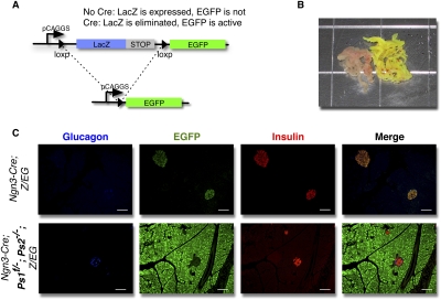
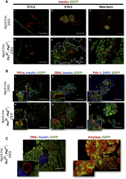
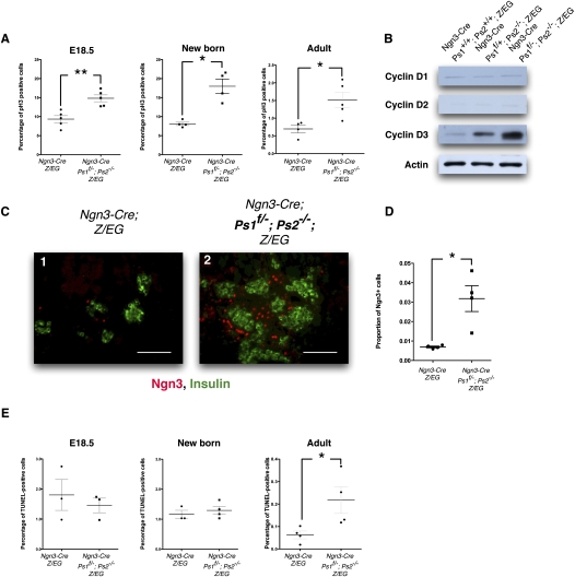
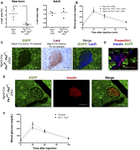
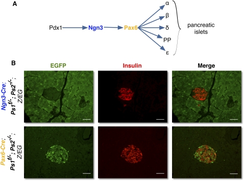
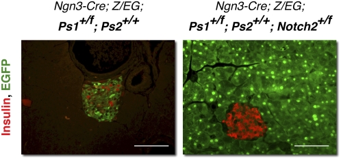
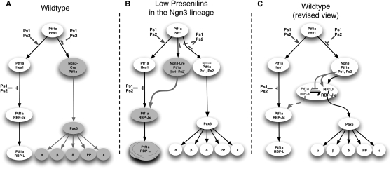
Canonical Notch signaling is thought to control the endocrine/exocrine decision in early pancreatic progenitors. Later, RBP-Jkappa interacts with Ptf1a and E12 to promote acinar differentiation. To examine the involvement of Notch signaling in selecting specific endocrine lineages, we deregulated this pathway by targeted deletion of presenilin1 and presenilin2, the catalytic core of gamma-secretase, in Ngn3- or Pax6-expressing endocrine progenitors. Surprisingly, whereas Pax6(+) progenitors were irreversibly committed to the endocrine fate, we discovered that Ngn3(+) progenitors were bipotential in vivo and in vitro. When presenilin amounts are limiting, Ngn3(+) progenitors default to an acinar fate; subsequently, they expand rapidly to form the bulk of the exocrine pancreas. gamma-Secretase inhibitors confirmed that enzymatic activity was required to block acinar fate selection by Ngn3 progenitors. Genetic interactions identified Notch2 as the substrate, and suggest that gamma-secretase and Notch2 act in a noncanonical titration mechanism to sequester RBP-Jkappa away from Ptf1a, thus securing selection of the endocrine fate by Ngn3 progenitors. These results revise the current view of pancreatic cell fate hierarchy, establish that Ngn3 is not in itself sufficient to commit cells to the endocrine fate in the presence of Ptf1a, reveal a noncanonical action for Notch2 protein in endocrine cell fate selection, and demonstrate that acquisition of an endocrine fate by Ngn3(+) progenitors is gamma-secretase-dependent until Pax6 expression begins.


Figures

Figure 1.
Figure 1.
Elimination of the Presenilins in vivo in Ngn3-progenitors. (A) Schematic representation of the Z/EG reporter. (B) Gross morphology of control (Ngn3; Z/EG) (left) and Presenilin-deficient (Ngn3-Cre; Ps1f/−; Ps2−/−; Z/EG) (right) pancreata of adult mice. (C) Immunostaining for glucagon (blue), EGFP (green), and insulin (red) in Ngn3; Z/EG (top) and Presenilin-deficient Ngn3-Cre; Ps1f/−; Ps2−/−; Z/EG (bottom) adult mice. Bars, 50 μm.
Figure 2.
Figure 2.
Presenilin-deficient pancreatic progenitors increase in numbers late during embryonic development. (A) Immunostaining for insulin (red) and EGFP (green) at E14.0 (left) and E18.5 (middle) and in the newborn (right) in control (Ngn3-Cre; Z/EG) (top) and presenilin-deficient (Ngn3-Cre; Ps1f/−; Ps2−/−; Z/EG) (bottom) mice. EGFP-positive cells are found outside of the endocrine compartment in Presenilin mice throughout development and expand around birth. (B) Coimmunostaining for Ptf1a (red), insulin (blue), and EGFP (green) (middle); DBA (red), insulin (blue), and EGFP (green) (right panel); or Pdx1 (red) DAPI (blue), and EGFP (green) (left panel) in sections of E18.5 control (Ngn3-Cre; Z/EG) (top) and Presenilin-deficient (Ngn3-Cre; Ps1f/−; Ps2−/−; Z/EG) (bottom) mice. Pslow, nonendocrine cells still express Pdx1, Ptf1a, and DBA at E18.5. (C) Coimmunostaining for DBA (red), Insulin (blue), and EGFP (green) (left panel), or Amylase (red) and EGFP (green) (right panel). Pslow cells exclusively express Amylase in the adult. Normal, EGFP-negative acinar cells form clusters n the periphery of the tissue. Bars, 50 μm.
Figure 3.
Figure 3.
Presenilin-deficient Ngn3-derived progenitors proliferate at a sustained rate and turn over rapidly in the adult. (A) Quantification of the percentage of Phospho-Histone3-positive cells in control and Presenilin-deficient mice at E18.5 (n = 25), newborn mice (n = 24), and adult mice (n = 25). More than 3900 nuclei were counted for each mouse. Presenilin-deficient mice display an increased proliferation rate at all stages. Gray bars represent SEM; (*) P < 0.05; (**) P < 0.01. (B) Western-blot analysis for the expression of Cyclins D1, D2, and D3 in total pancreatic extracts of control (Ngn3-Cre; Z/EG) and Presenilin-deficient (Ngn3-Cre; Ps1+/f; Ps2−/−; Z/EG and Ngn3-Cre; Ps1f/−; Ps2−/−; Z/EG). The immunoblots are normalized over actin and indicate that Cyclin D3 increased concomitantly with the reduction of Presenilins. (C) Expression of Ngn3 around forming islets at E18.5 in control (panel 1) and Presenilin-deficient (panel 2) mice. Ngn3 is stained in red and insulin is stained in green. Bars, 50 μm. (D) Overall proportion of Ngn3+ cells is represented on the right. Gray bars represent SEM; P < 0.05. (E) Quantification of the percentage of TUNEL-positive cells in control and Presenilin-deficient mice at E18.5 (n = 23), newborn mice (n = 24), and adult mice (n = 24); >3400 nuclei were counted for each mouse. The apoptosis rate is indistinguishable between control and Presenilin-deficient mice at E18.35 and at birth, but increases in Presenilin mice in the adult. Gray bars represent SEM; P < 0.05.
Figure 4.
Figure 4.
Ngn3-Cre Ps1f/−; Ps2−/−; Z/EG compensate and form normal islets through mosaicism. (A) Morphometric measurement of the β-cell mass of controls (Ngn3-Cre; Z/EG) and Presenilin-deficient (Ngn3-Cre Ps1f/−; Ps2−/−; Z/EG) mice at birth (left) and in the adult (right). The measurements are expressed as a percentage of staining for β cells over the entire area for the pancreas, and is further normalized to the total mass of the pancreas in the adult (n = 23–4). Total pancreas weights are similar between groups. Gray bars represent the SEM for each group. The β-cell mass is significantly reduced in Presenilin-deficient mice at birth, but increases to reach that of the control mice in the adult. (B) Intraperitoneal glucose tolerance test on control (Ngn3-Cre; Z/EG) and Presenilin-deficient (Ngn3-Cre; Ps1+/f; Ps2−/−; Z/EG and Ngn3-Cre Ps1f/−; Ps2−/−; Z/EG) adult mice, indicating no difference in glucose response between the two groups. (C) Endogenous EGFP expression (green) (left) and LacZ staining (red, with hematoxylin counterstain in blue) (right) on Ngn3-Cre; Ps1f/−; Ps2−/−; Z/EG mice. The fields are merged in false color (EGFP in green, LacZ in blue) on the right. (D) Endogenous EGFP expression (green) and immunostaining for Presenilin1 (red) and insulin (blue) on Ngn3-Cre; Ps1+/f; Ps2−/−; Z/EG mice. Presenilin-positive cells are EGFP-negative. (E) Immunostaining for insulin (red) and EGFP (green) in pancreatic sections from Ngn3-Cre; Ps1+/−; Ps2−/−; Z/EG mice. Bars, 50 μm. (F) Glucose tolerance tests on control (wild-type) and Ps1+/−; Ps2−/− mice. n = 25 for each group.
Figure 5.
Figure 5.
Presenilins control the endocrine fate through a γ-secretase cleavage and Notch activation. (A) Cultures of E13.0 pancreatic rudiments from Ngn3; Z/EG rudiments with (bottom) or without (top) a γ-secretase inhibitor (DAPT) for 7 d. Immunostaining for insulin (blue), EGFP (green), and Amylase (red). Insets present higher magnifications for Amylase+, EGFP+ areas, indicating that the Presenilin-deficient phenotype can be recapitulated through γ-secretase inhibition. Bars, 50 μm. (B) BrdU incorporation rate in E13.0 pancreatic rudiments kept in culture for 7 d with (right) or without (left) DAPT. Four rudiments were used for each group, and 3500–4000 cells were counted for each rudiment. Gray bar represents the SEM for each group; P < 0.05. (C) BrdU incorporation rate in E13.0 pancreatic rudiments kept in culture for 8 d in control conditions, or for 7 d in control condition (left, n = 24), then with DAPT for an additional day (right, n = 25). Approximately 4000 cells were counted for each rudiment. Gray bar represents the SEM for each group; P < 0.05.
Figure 6.
Figure 6.
Presenilins are not required for endocrine determination after Pax6 expression. (A) Schematic representation of the differentiation pathway of the pancreatic endocrine progenitors. (B) Immunostaining for insulin (red) and EGFP (green) on Ngn3-Cre; Ps1f/−; Ps2−/−; Z/EG (top) and Pax6-Cre; Ps1f/−; Ps2−/−; Z/EG (bottom) mice. Bars:,50 μm.
Figure 7.
Figure 7.
Endocrine fate is controlled by Notch2. Immunostaining for insulin (red) and EGFP (green) in phenotypically wild-type Ngn3-Cre, Ps1+/f; Ps2+/+; Z/EG mice and in Ngn3-Cre, Ps1+/f; Ps2+/+; Notch2+/f; Z/EG mice indicates that eliminating one copy of notch2 in Ngn3 progenitors can enhance the reduction in Presenilins and recapitulate the Pslow phenotype. Bars, 50 μm.
Figure 8.
Figure 8.
Presenilins are involved in multiple steps of pancreatic development. (A) Representation of the previous view of the control of the fate of the pancreatic progenitors. (B) Representation of the alteration of fate in low abundance of the Presenilins in Ngn3 progenitors. (C) Proposed revised view of the control of the fate of the endocrine progenitors.

Similar articles

Cited by

References

    1. Apelqvist Å, Li H, Sommer L, Beatus P, Anderson DJ, Honjo T, Hrabe de Angelis M, Lendahl U, Edlund H. Notch signalling controls pancreatic cell differentiation. Nature. 1999;400:877–881. - PubMed
    1. Baki L, Shioi J, Wen P, Shao Z, Schwarzman A, Gama-Sosa M, Neve R, Robakis NK. PS1 activates PI3K thus inhibiting GSK-3 activity and tau overphosphorylation: Effects of FAD mutations. EMBO J. 2004;23:2586–2596. - PMC - PubMed
    1. Beres TM, Masui T, Swift GH, Shi L, Henke RM, Macdonald RJ. PTF1 is an organ-specific and Notch-independent basic helix–loop–helix complex containing the mammalian suppressor of hairless (RBP-J) or its paralogue, RBP-L. Mol Cell Biol. 2006;26:117–130. - PMC - PubMed
    1. Bernal-Mizrachi E, Wen W, Stahlhut S, Welling CM, Permutt MA. Islet β cell expression of constitutively active Akt1/PKB α induces striking hypertrophy, hyperplasia, and hyperinsulinemia. J Clin Invest. 2001;108:1631–1638. - PMC - PubMed
    1. Cheng HT, Miner JH, Lin M, Tansey MG, Roth K, Kopan R. γ-Secretase activity is dispensable for mesenchyme-to-epithelium transition but required for podocyte and proximal tubule formation in developing mouse kidney. Development. 2003;130:5031–5042. - PubMed

Publication types

Substances