

Imaging features of hepatic angiomyolipomas on real-time contrast-enhanced ultrasound
- PMID: 19723766
- PMCID: PMC3473573
- DOI: 10.1259/bjr/81174247
Imaging features of hepatic angiomyolipomas on real-time contrast-enhanced ultrasound
Abstract
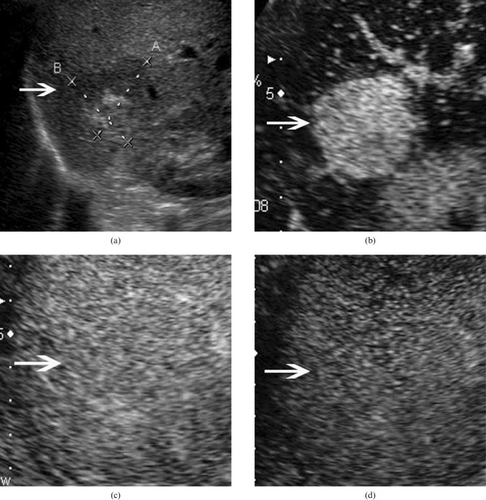
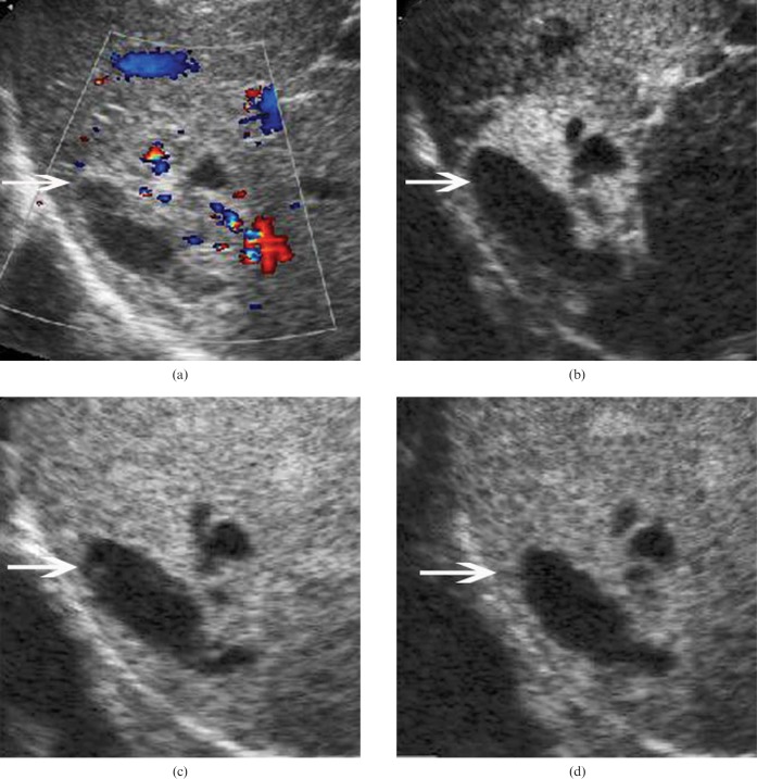
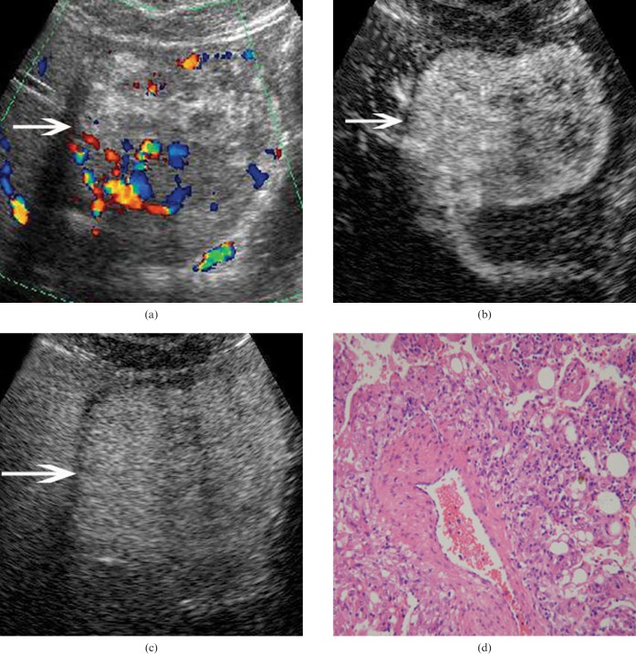
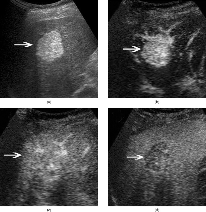
The aim of this study was to evaluate the imaging features of hepatic angiomyolipoma (AML) on contrast-enhanced ultrasound (CEUS). The imaging features of 12 pathologically proven hepatic AML lesions in 10 patients who had undergone baseline ultrasound (BUS) and CEUS examinations were evaluated retrospectively. The enhancement extent, pattern and dynamic change, along with the enhancement process, on CEUS were analysed. The diagnostic results of BUS and CEUS before pathological examination were also recorded. The results showed that 75% (9/12) of the AML lesions exhibited mixed echogenicity on BUS and most showed remarkable hyperechogenicity in combination with a hypoechoic or anechoic portion. Arterial flow signals were detected in 75% (9/12) of the lesions on colour Doppler imaging. On CEUS, 66.7% (n = 8) of the 12 lesions exhibited hyperenhancement in the arterial phase, slight hyperenhancement (n = 2) or isoenhancement (n = 6) in the portal phase, and slight hyperenhancement (n = 1) or isoenhancement (n = 7) in the late phase. Three (25%) lesions exhibited hyperenhancement in the arterial phase and hypoenhancement in both portal and late phases. One (8.3%) lesion exhibited hypoenhancement throughout the CEUS process. Before pathological examination with BUS, only 3 (25%) lesions were correctly diagnosed as hepatic AML. Conversely, on CEUS, correct diagnoses were made for 66.8% (8/12) of hepatic AMLs. Therefore, arterial hyperenhancement and subsequent sustained enhancement on CEUS were found in the majority of hepatic AMLs. The combination of BUS and CEUS leads to the correct diagnosis in the majority of hepatic AMLs, and is higher than the success rate achieved by BUS alone.
Figures
Similar articles
-
Real-time contrast-enhanced ultrasonography of resected and immunohistochemically proven hepatic angiomyolipomas.Abdom Imaging. 2010 Dec;35(6):676-82. doi: 10.1007/s00261-009-9592-x. Abdom Imaging. 2010. PMID: 20020286
-
Contrast-enhanced ultrasound of hepatocarcinogenesis in liver cirrhosis.Chin Med J (Engl). 2012 Sep;125(17):3104-9. Chin Med J (Engl). 2012. PMID: 22932189
-
Hepatic angiomyolipomas: ultrasonic characteristics of 25 patients from a single center.Ultrasound Med Biol. 2015 Feb;41(2):393-400. doi: 10.1016/j.ultrasmedbio.2014.09.014. Epub 2014 Dec 23. Ultrasound Med Biol. 2015. PMID: 25542497
-
Washout on Contrast-Enhanced Ultrasound of Benign Focal Liver Lesions-A Review on Its Frequency and Possible Causes.Diagnostics (Basel). 2025 Apr 14;15(8):998. doi: 10.3390/diagnostics15080998. Diagnostics (Basel). 2025. PMID: 40310346 Free PMC article. Review.
-
Myomatous hepatic angiomyolipoma: imaging findings in 14 cases with radiological-pathological correlation and review of the literature.Br J Radiol. 2014 Jun;87(1038):20130712. doi: 10.1259/bjr.20130712. Epub 2014 Mar 27. Br J Radiol. 2014. PMID: 24670055 Free PMC article. Review.
Cited by
-
Shear-wave elastography combined with contrast-enhanced ultrasound algorithm for noninvasive characterization of focal liver lesions.Radiol Med. 2023 Jan;128(1):6-15. doi: 10.1007/s11547-022-01575-5. Epub 2022 Dec 16. Radiol Med. 2023. PMID: 36525179
-
Hepatic monotypic epithelioid angiomyolipoma with concomitant hepatocellular carcinoma.Int J Clin Exp Pathol. 2019 Apr 1;12(4):1399-1405. eCollection 2019. Int J Clin Exp Pathol. 2019. PMID: 31933955 Free PMC article.
-
Hepatic perivascular epithelioid cell tumors: The importance of preoperative diagnosis.World J Gastroenterol. 2024 Apr 7;30(13):1926-1933. doi: 10.3748/wjg.v30.i13.1926. World J Gastroenterol. 2024. PMID: 38659487 Free PMC article. Review.
-
Hepatic Angiomyolipoma Staining in the Post-vascular Phase of Contrast-enhanced Ultrasound Due to the Presence of Macrophages.Intern Med. 2018 May 1;57(9):1247-1251. doi: 10.2169/internalmedicine.9697-17. Epub 2017 Dec 27. Intern Med. 2018. PMID: 29279500 Free PMC article.
-
Contrast-enhanced ultrasound features of hepatic angiomyolipoma: comparison with AFP-negative and non-viral hepatocellular carcinoma.Ultrasound Int Open. 2024 Jun 14;10:a23186654. doi: 10.1055/a-2318-6654. eCollection 2024. Ultrasound Int Open. 2024. PMID: 39411752 Free PMC article.
References
-
- Nonomura A, Mizukami Y, Kadoya M. Angiomyolipoma of the liver: a collective review. J Gastroenterol 1994;29:95–105 - PubMed
-
- Tsui WM, Colombari R, Portmann BC, Bonetti F, Thung SN, Ferrell LD, et al. Hepatic angiomyolipoma: a clinicopathologic study of 30 cases and delineation of unusual morphologic variants. Am J Surg Pathol 1999;23:34–48 - PubMed
-
- Horton KM, Bluemke DA, Hruban RH, Soyer P, Fishman EK. CT and MR imaging of benign hepatic and biliary tumors. Radiographics 1999;19:431–51 - PubMed
-
- Rettenbacher T. Focal liver lesions: role of contrast-enhanced ultrasound. Eur J Radiol 2007;64:173–82 - PubMed
-
- Albrecht T, Blomley M, Bolondi L, Claudon M, Correas JM, Cosgrove D, et al. Guidelines for the use of contrast agents in ultrasound. January 2004. Ultraschall Med 2004;25:249–56 - PubMed
Publication types
MeSH terms
Substances
LinkOut - more resources
Full Text Sources
Medical
