The contribution of 3D-CISS and contrast-enhanced MR cisternography in detecting cerebrospinal fluid leak in patients with rhinorrhoea
- PMID: 19723768
- PMCID: PMC3473545
- DOI: 10.1259/bjr/56838652
The contribution of 3D-CISS and contrast-enhanced MR cisternography in detecting cerebrospinal fluid leak in patients with rhinorrhoea
Abstract
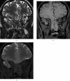
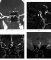
The aim of this prospective study was to evaluate the value of unenhanced (three-dimensional constructive interference in steady state (3D-CISS)) and contrast-enhanced MR cisternography (CE-MRC) in detecting the localisation of cerebrospinal fluid (CSF) leak in patients with rhinorrhoea. 17 patients with active or suspected CSF rhinorrhoea were included in the study. 3D-CISS sequences in coronal and sagittal planes and fat-suppressed T1-weighted spin-echo sequences in three planes before and after intrathecal contrast media administration were obtained. Images were obtained of the cribriform plate and sphenoid sinus. In addition, high-resolution CT (HRCT) was performed in order to evaluate the bony elements. The leak was present in 9/17 patients with 3D-CISS and 10/17 patients with CE-MRC. The leak from the cribriform plate to the nasal cavity in six patients and from the sphenoid sinus in four patients was nicely shown by CE-MRC. Eight of those patients were surgically treated, but spontaneous regression of the symptoms in two precluded any intervention. The leak localisations shown with CE-MRC were fully compatible with surgical results. The sensitivities of HRCT, 3D-CISS and CE-MRC for showing CSF leakage were 88%, 76% and 100%, respectively. In conclusion, 3D-CISS is a non-invasive and reliable technique, and should be the first-choice method to localise CSF leak. CE-MRC is helpful in conditions when there is no leak or in complicated cases with a positive beta2-transferrin measurement.
Figures


Similar articles
-
3D steady-state MR cisternography in CSF rhinorrhoea.Acta Radiol. 2001 Nov;42(6):582-4. doi: 10.1080/028418501127347412. Acta Radiol. 2001. PMID: 11736705 Clinical Trial.
-
Intrathecal gadolinium-enhanced magnetic resonance cisternography in cerebrospinal fluid rhinorrhea: road ahead?J Neurotrauma. 2007 Oct;24(10):1570-5. doi: 10.1089/neu.2007.0326. J Neurotrauma. 2007. PMID: 17970620
-
Phase-contrast MRI and 3D-CISS versus contrast-enhanced MR cisternography for the detection of spontaneous third ventriculostomy.J Neuroradiol. 2011 May;38(2):98-104. doi: 10.1016/j.neurad.2010.03.006. Epub 2010 Jun 7. J Neuroradiol. 2011. PMID: 20627312
-
Traumatic, iatrogenic, and spontaneous cerebrospinal fluid (CSF) leak: endoscopic repair.B-ENT. 2011;7 Suppl 17:47-60. B-ENT. 2011. PMID: 22338375 Review.
-
Intrathecal Contrast-enhanced Computed Tomography and MR Cisternography for Skull Base Cerebrospinal Fluid Leaks and Other Intracranial Applications.Neuroimaging Clin N Am. 2025 Feb;35(1):105-121. doi: 10.1016/j.nic.2024.08.025. Epub 2024 Sep 24. Neuroimaging Clin N Am. 2025. PMID: 39521519 Review.
Cited by
-
Diagnosis and Localization of Cerebrospinal Fluid Rhinorrhea: A Systematic Review.Am J Rhinol Allergy. 2022 May;36(3):397-406. doi: 10.1177/19458924211060918. Epub 2021 Nov 30. Am J Rhinol Allergy. 2022. PMID: 34846218 Free PMC article.
-
Non-Invasive and Minimally Invasive Imaging Evaluation of CSF Rhinorrhoea - a Retrospective Study with Review of Literature.Pol J Radiol. 2016 Feb 29;81:80-5. doi: 10.12659/PJR.895698. eCollection 2016. Pol J Radiol. 2016. PMID: 26985244 Free PMC article.
-
Imaging of cerebrospinal fluid leakage from the cribriform plate.Radiol Case Rep. 2025 Jan 20;20(4):1920-1924. doi: 10.1016/j.radcr.2025.01.004. eCollection 2025 Apr. Radiol Case Rep. 2025. PMID: 39897741 Free PMC article.
-
Pseudo-Cerebrospinal Fluid Leaks of the Anterior Skull Base: Algorithm for Diagnosis and Management.J Neurol Surg B Skull Base. 2021 Jun;82(3):351-356. doi: 10.1055/s-0039-3399519. Epub 2019 Nov 8. J Neurol Surg B Skull Base. 2021. PMID: 34026412 Free PMC article.
-
Recurrent Community-Acquired Bacterial Meningitis in Adults.Clin Infect Dis. 2021 Nov 2;73(9):e2545-e2551. doi: 10.1093/cid/ciaa1623. Clin Infect Dis. 2021. PMID: 33751028 Free PMC article.
References
-
- Sirikici A, Bayazit Y, Bayram M, KervanciogluR MRI Findings Simulating CSF Leakage on Routine Imaging. Turk J Diagn Intervent Radiol 2000;6:283–6
-
- Aydin K, Terzibasioglu E, Sencer S, Sencer A, Suoglu Y, Karasu A, Kiris T, Turantan M. Localization of cerebrospinal fluid leaks by gadolinium-enhanced magnetic resonance cisternography: a 5-year single-center experience. Neurosurgery 2008;62:584–9 - PubMed
-
- Jayakumar PN, Kovoor JME, Srikanth SG, Praharaj SS. 3D Steady-State MR Cisternography in CSF Rhinorrhoea. Acta Radiol 2001;42:582–4 - PubMed
MeSH terms
Substances
LinkOut - more resources
Full Text Sources
Medical

