. 2009 Oct 29;114(18):3928-34.
doi: 10.1182/blood-2009-06-230086. Epub 2009 Sep 1.

The water channel aquaporin-1 partitions into exosomes during reticulocyte maturation: implication for the regulation of cell volume

Affiliations

The water channel aquaporin-1 partitions into exosomes during reticulocyte maturation: implication for the regulation of cell volume

Lionel Blanc et al. Blood. .

Abstract

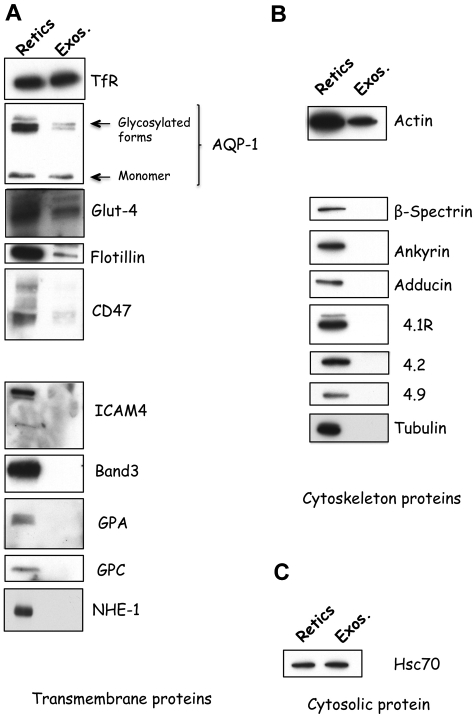
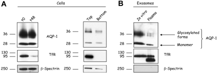
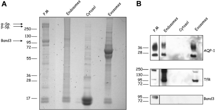
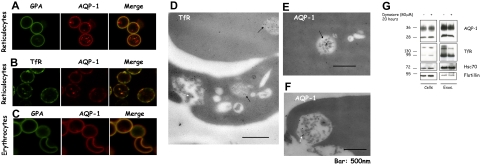
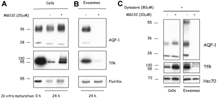
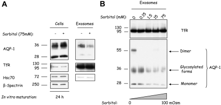
Aquaporin-1 (AQP-1), the universal water channel, is responsible for rapid response of cell volume to changes in plasma tonicity. In the membrane of the red cell the concentration of the protein is tightly controlled. Here, we show that AQP-1 is partially lost during in vitro maturation of mouse reticulocytes and that it is associated with exosomes, released throughout this process. AQP-1 in young reticulocytes localizes to the plasma membrane and also in endosomal compartments and exosomes, formed both in vitro and in vivo. During maturation a part of the total pool of AQP-1 is differentially sorted and released via the exosomal pathway. A proteasome inhibitor, MG132, suppresses secretion of AQP-1, implying that ubiquitination is a sorting signal for its release. We further show that modulation of medium tonicity in vitro regulates the secretion of AQP-1, thus showing that extracellular osmotic conditions can drive sorting of selected proteins by the exosomal pathway. These results lead us to suggest that AQP-1 sorting into exosomes may be the mechanism by which the reticulocyte adapts to environmental changes during its maturation.


Figures

Figure 1
Figure 1
Specificity of protein sorting into exosomes during reticulocyte maturation. Reticulocytes (Retics; 106 cells; left lanes) and exosomes (Exos.; 50 μg protein; right lanes) were assessed for the presence of transmembrane (A), cytoskeletal (B), or cytosolic (C) proteins by SDS-PAGE and immunoblotting for the proteins, as indicated on the right.
Figure 2
Figure 2
AQP-1 is released in association with exosomes during in vitro maturation of reticulocytes and in vivo. (A) Reticulocytes (106 cells) before (t0) and after 48 hours (t48) in vitro maturation (left); top and bottom fractions were collected after cell separation on a Percoll gradient (right) were subjected to SDS-PAGE, followed by immunoblotting for the proteins indicated. (B) Exosomes (50 μg protein) released during in vitro maturation (left lane) or isolated directly from the plasma of anemic mice (right lane) were subjected to SDS-PAGE, followed by immunoblotting for the proteins indicated.
Figure 3
Figure 3
Subcellular localization of reticulocyte AQP-1. Constituents of mouse reticulocytes were fractionated after lysis as described in “Methods.” The various fractions (50 μg protein), plasma membrane, endosomes, cytosol, and exosomes were separated by SDS-PAGE and (A) stained with Coomassie Blue or (B) immunoblotted for the proteins indicated. Arrows indicate cytoskeleton proteins present in the mature red cell plasma membrane. Blots of plasma membrane fraction were exposed for a shorter period of time than those of the other 3 samples as indicated by the solid lines.
Figure 4
Figure 4
A part of the pool of AQP-1 colocalizes with the TfR and is found in multivesicular endosomes in reticulocytes. After purification by Percoll gradient, reticulocytes (A-B) and red blood cells (C) were fixed with acrolein and deposited on cover slips pretreated with poly-l-lysine. After permeabilization (Triton X-100), the cells were immunostained for GPA (green) and AQP-1 (red) (A,C), or TfR (green) and AQP-1 (red) (B). The slides were examined in a confocal microscope (Zeiss LSM 510 META camera, Zeiss Plan Apochromat 100×/1.4 NA oil objective, scan zoom 2.0) and pictures acquired and processed with Zeiss Laser Scanning Microscope LSM 510 software, Version 3.2 SP2. For visualization of TfR and AQP-1 in multivesicular endosomes and exosomes, reticulocytes were fixed with a mixture of paraformaldehyde, glutaraldehyde, and sucrose. After dehydration and embedding, thin sections of cells were mounted on nickel grids, blocked, and then immunostained for the TfR (D) or AQP-1 (E-F). Sections were examined in a Philips-410 electron microscope equipped with AMT XR41 Biological 2k side-mounted CCD camera, and images were acquired with AMT600 software and analyzed with Photoshop CS (Adobe Systems). After treatment with dynasore 80 μM or the solvent, DMSO, alone for 20 hours (G), 106 cells (left) and exosomes, resuspended in the same volume of Laemmli buffer, (right) were examined by SDS-PAGE and immunoblotting for the proteins indicated.
Figure 5
Figure 5
Inhibition of AQP-1 trafficking and sorting by MG132. (A) Freshly obtained mouse reticulocytes (0 hour; left lane) were cultured for 24 hours with the proteasome inhibitor MG132 (20 μM; 24 hours; right lane). Freshly obtained mouse reticulocytes were also cultured with an equivalent volume of the solvent, DMSO (24 hours; middle lane). (B) Exosomes were isolated from the culture medium after 24 hours and resuspended in the same volume of Laemmli buffer. Cellular and exosomal proteins were separated by SDS-PAGE and immunoblotted for the proteins indicated on the right. In each lane, 106 cells were loaded. (C) After inhibition of endocytosis, the cells were allowed to mature in the presence or absence of MG132 for 24 hours. Exosomes were then collected, and the compositions of both cells and the vesicles released from them were analyzed by SDS-PAGE and immunoblotted for the same proteins.
Figure 6
Figure 6
Hypertonic stress progressively inhibits AQP-1 sorting into exosomes. (A) Mouse reticulocytes were cultured for 24 hours with or without sorbitol 75 mM. Exosomes were then collected, and both cells (106) and exosomes (resuspended in the same volume of Laemmli buffer) were examined by SDS-PAGE and immunoblotted for the proteins indicated on the right. (B) Mouse reticulocytes were cultured for 24 hours, with increasing concentrations of sorbitol, and exosomes were collected, resuspended in the same volume of Laemmli buffer, and examined by SDS-PAGE and immunoblotted for TfR and AQP-1.
Figure 7
Figure 7
Proposed model of AQP-1 sorting into exosomes during reticulocyte maturation. Under physiologic conditions (A), AQP-1 levels are regulated through the exosomal pathway. During osmotic stress, by contrast (B), AQP-1 secretion is inhibited, probably by regulation of its ubiquitination or by phosphorylation. It is consequently rerouted from the secretion pathway into the recycling pathway, thereby augmenting the AQP-1 concentration on the plasma membrane, and thus assisting the cell to nullify the osmotic shock. TfR trafficking, by contrast, is not affected by the osmotic disturbance and continues to be sorted and secreted through the exosomal pathway.

References

    1. Mel HC, Prenant M, Mohandas N. Reticulocyte motility and form: studies on maturation and classification. Blood. 1977;49(6):1001–1009. - PubMed
    1. Chasis JA, Mohandas N. Erythroblastic islands: niches for erythropoiesis. Blood. 2008;112(3):470–478. - PMC - PubMed
    1. Gronowicz G, Swift H, Steck TL. Maturation of the reticulocyte in vitro. J Cell Sci. 1984;71(1):177–197. - PubMed
    1. Johnstone RM. The Jeanne Manery-Fisher Memorial Lecture 1991. Maturation of reticulocytes: formation of exosomes as a mechanism for shedding membrane proteins. Biochem Cell Biol. 1992;70(3):179–190. - PubMed
    1. Rieu S, Geminard C, Rabesandratana H, Sainte-Marie J, Vidal M. Exosomes released during reticulocyte maturation bind to fibronectin via integrin alpha4beta1. Eur J Biochem. 2000;267(2):583–590. - PubMed

Publication types