. 2009 Dec;29(12):1955-67.
doi: 10.1038/jcbfm.2009.177. Epub 2009 Sep 2.

Cerebrovascular responses to insulin in rats

Affiliations

Cerebrovascular responses to insulin in rats

Prasad Venkateswera Gurunath Katakam et al. J Cereb Blood Flow Metab. 2009 Dec.

Abstract

Effects of insulin on cerebral arteries have never been examined. Therefore, we determined cerebrovascular actions of insulin in rats. Both PCR and immunoblot studies identified insulin receptor expression in cerebral arteries and in cultured cerebral microvascular endothelial cells (CMVECs). Diameter measurements (% change) of isolated rat cerebral arteries showed a biphasic dose response to insulin with an initial vasoconstriction at 0.1 ng/mL (-9.7%+/-1.6%), followed by vasodilation at 1 to 100 ng/mL (31.9%+/-1.4%). Insulin also increased cortical blood flow in vivo (30%+/-8% at 120 ng/mL) when applied topically. Removal of reactive oxygen species (ROS) abolished the vasoconstriction to insulin. Endothelial denudation, inhibition of K(+) channels, and nitric oxide (NO) synthase, all diminished insulin-induced vasodilation. Inhibition of cytochrome P450 enhanced vasodilation in endothelium-intact arteries, but promoted vasoconstriction after endothelial denudation. Inhibition of cyclooxygenase abolished vasoconstriction and enhanced vasodilation to insulin in all arteries. Inhibition of endothelin type A receptors enhanced vasodilation, whereas endothelin type B receptor blockade diminished vasodilation. Insulin treatment in vitro increased Akt phosphorylation in cerebral arteries and CMVECs. Fluorescence studies of CMVECs showed that insulin increased intracellular calcium and enhanced the generation of NO and ROS. Thus, cerebrovascular responses to insulin were mediated by complex mechanisms originating in both the endothelium and smooth muscle.


Conflict of interest statement

Conflict of interest

The authors declare no conflict of interest.

Figures
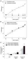
Figure 1
Figure 1
Effects of insulin on the diameter of rat cerebral arteries and cortical blood flow. Vascular responses to insulin in grouped rat cerebral arteries with intact endothelium and endothelium denuded are shown in (A). Responses to insulin in endothelium intact middle and posterior cerebral arteries have been presented separately in (B). Data are mean ± s.e.m. of 12 to 21 experiments. (*) indicates significant difference with regard to insulin response in endothelium intact arteries (P < 0.05). (C) Bar graph of average % changes of cortical blood flow (CoBF) at the first, sixth and tenth minutes of artificial cerebrospinal fluid (aCSF) and insulin (12 and 120 ng/mL) application. The % change in CoBF data is presented as mean ± s.e.m. (n=4). (*) indicates significant increase in CoBF compared with the % change in CoBF following the application of vehicle (P < 0.05). Insulin at high concentration (120 ng/mL) significantly increased the CoBF when compared with the vehicle (aCSF) and the low-dose insulin.
Figure 2
Figure 2
Role of NOS, COX, cytochrome P450, K+ channels, endothelium and ROS in cerebrovascular responses to insulin. Vascular responses to insulin in rat cerebral arteries with intact endothelium, in the presence and absence of indomethacin, miconazole, and KCl are shown in (A). In (B), vascular responses to insulin in endothelium-denuded arteries in the presence and absence of indomethacin and miconazole are shown. Insulin responses in the presence and absence of L-NAME, 7-nitroindazole, and L-NAME + indomethacin combination are shown in (C). (D) Shows the responses to insulin in the absence and presence of MnTBAP and apocynin. Baseline response indicates insulin response alone in endothelium-intact arteries in the absence of any drugs. Data are mean ± s.e.m. of 5 to 21 experiments. (*) Indicates significant difference with regard to insulin response alone (P < 0.05).
Figure 3
Figure 3
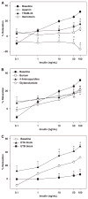
Contribution of K+ channels and endothelin receptors to cerebrovascular responses to insulin. Vascular responses to insulin in rat cerebral arteries in the presence and absence of iberiotoxin, apamin, and TRAM-34 are shown in (A). Insulin responses in the absence and presence of barium, 4-AP, and glybenclamide are shown in (B). In (C), responses to insulin in the presence and absence of endothelin type A (ETA) inhibitor BQ-123 and endothelin type B (ETB) inhibitor, BQ-788, are shown. Baseline response indicates insulin response alone in endothelium intact arteries in the absence of any drug. Data are mean ± s.e.m. of 5 to 21 experiments. (*) indicates significant difference with respect to insulin response alone (P<0.05).
Figure 4
Figure 4
Effect of insulin on intracellular calcium dynamics and generation of NO and ROS in cerebral microvascular endothelial cells. Fluorescence measurements of calcium influx and generation of NO and ROS in the cultured rat CMVECs. (A) Representative fluorescence images of CMVECs loaded with the calcium sensitive fluoroprobe, fluo-4 AM, following the administration of vehicle (top) and 100 ng/mL insulin (bottom). The group data of the % change in the intensity of fluo-4 AM fluorescence from baseline in response to insulin compared with vehicle is shown as a bar graph in (B). (C) Representative fluorescence images of CMVECs loaded with the NO-sensitive fluoroprobe, DAF-FM diacetate, following the administration of vehicle (top) and 100 ng/mL insulin (bottom) alone or in the presence of NOS inhibitor, L-NAME. The group data of the % change in the intensity of fluo-4 AM fluorescence from baseline in response to insulin compared with vehicle in the presence and absence of L-NAME is shown as a bar graph in (D). (E) Representative fluorescence images of CMVECs loaded with the ROS-sensitive dye, hydroethidine (HEt), in the presence of vehicle (top) or 100 ng/mL insulin (bottom). The group data of the % increase in the intensity of HEt fluorescence in response to insulin compared with vehicle is shown as a bar graph in (F). (*) indicates significant difference in fluorescence intensity in response to insulin compared with the vehicle (P<0.05).
Figure 5
Figure 5
Expression of insulin receptor β and the effect of insulin on AKt phosphorylation in rat cerebral arteries and cerebral microvascular endothelial cells. RT-PCR analysis was performed to identify the mRNA of insulin receptor-β subunit. (A) Shows the bands representing the PCR products of insulin receptor-β in isolated rat cerebral arteries, CMVECs, and the liver from Sprague–Dawley rats. (B) Shows the immunobands identifying the insulin receptor-β subunit in homogenates of cerebral arteries, CMVECs, and the liver from Sprague–Dawley rats. (C) Shows the representative western blots of total and phosphorylated Akt protein along with β-actin in the homogenates of cerebral arteries (top) from three separate Sprague–Dawley rats and CMVECs (2 groups of rats, bottom) treated with 100 ng/mL insulin (+) and untreated controls (−). The ratio of the intensity of immunobands of phosphorylated Akt and total Akt normalized to β-actin immunobands in the presence and absence of insulin is represented as a bar graph in (D). (*) Indicates significant difference with regard to untreated cerebral arteries (P<0.05).
Figure 6
Figure 6
A schematic of the mechanisms underlying cerebrovascular actions of insulin in rats. Insulin-induced vasodilation is primarily endothelium-dependent, mediated by prostacyclin (PGI2), nitric oxide (NO), and endothelium-derived hyperpolarizing factor (EDHF). Elevation of endothelial calcium [Ca2+]i leads to activation of endothelial NO synthase (eNOS), cyclooxygenase, and EDHF pathway, although insulin-induced activation of eNOS is primarily calcium-independent. Calcium-activated K+ channels (SKCa, small; IKCa, intermediate; BKCa, large-conductance), myoendothelial gap junctions and inwardly-rectifying K+ channels (Kir) have all been implicated in the vascular hyperpolarization-mediated vasodilation (EDHF mechanism). In addition, NO and PGI2 activate guanylate and adenylate cyclase to produce cGMP and cAMP in vascular smooth muscle (VSM) cells, respectively, leading to activation of ATP-dependent K+ channels and BKCa. The subsequent K+ efflux and hyperpolarization promotes decrease in [Ca2+]i of VSM cells leading to vasodilation. In addition, cGMP and cAMP induce vasodilation by calcium-independent mechanism. Insulin-induced vasoconstriction results from the production of endothelin, vasoconstrictor prostanoids by COX, arachidonic acid metabolites by CP450 or reactive oxygen species (ROS) by the entire vascular wall. ROS induce vasoconstriction by either decreasing the NO bioavailability or promoting direct vasoconstriction. Endothelin causes predominantly vasoconstriction by activation of type A receptors (ETA) in VSM cells, whereas type B receptor (ETB) activation contributes to endothelium-dependent vasodilation. The vasoconstrictor factors promote vasoconstriction by both calcium-dependent (increased [Ca2+]i) and independent mechanisms. Dotted arrows in the figure represent inhibition mechanisms.

References

    1. Brahler S, Kaistha A, Schmidt VJ, Wolfle SE, Busch C, Kaistha BP, Kacik M, Hasenau AL, Grgic I, Si H, Bond CT, Adelman JP, Wulff H, de Wit C, Hoyer J, Kohler R. Genetic deficit of SK3 and IK1 channels disrupts the endothelium-derived hyperpolarizing factor vasodilator pathway and causes hypertension. Circulation. 2009;119:2323–32. - PubMed
    1. Chen YL, Messina EJ. Dilation of isolated skeletal muscle arterioles by insulin is endothelium dependent and nitric oxide mediated. Am J Physiol. 1996;270:H2120–4. - PubMed
    1. Eichler I, Wibawa J, Grgic I, Knorr A, Brakemeier S, Pries AR, Hoyer J, Kohler R. Selective blockade of endothelial Ca2+-activated small- and intermediate-conductance K+-channels suppresses EDHF-mediated vasodilation. Br J Pharmacol. 2003;138:594–601. - PMC - PubMed
    1. Erdos B, Snipes JA, Miller AW, Busija DW. Cerebrovascular dysfunction in Zucker obese rats is mediated by oxidative stress and protein kinase C. Diabetes. 2004;53:1352–9. - PubMed
    1. Eringa EC, Stehouwer CD, Merlijn T, Westerhof N, Sipkema P. Physiological concentrations of insulin induce endothelin-mediated vasoconstriction during inhibition of NOS or PI3-kinase in skeletal muscle arterioles. Cardiovasc Res. 2002;56:464–71. - PubMed

Publication types

MeSH terms