

Primary cardiac angiosarcoma: a fatal disease
- PMID: 19724650
- PMCID: PMC2731464
- DOI: 10.1155/2009/591512
Primary cardiac angiosarcoma: a fatal disease
Abstract
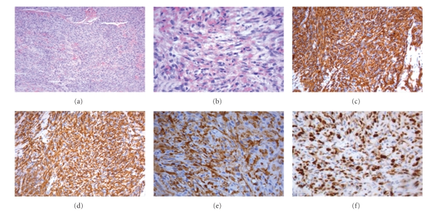
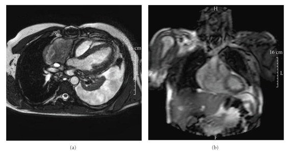
A 42-year-old man with a cardiac tamponade underwent an urgent pericardiotomy that showed tumoral tissue, covering the surface of the right atrium. The tumor was then partially excised, and the histological examination revealed the presence of a moderately-differentiated angiosarcoma. The patient was then referred to the oncology unit and scheduled for a chemotherapy schedule including Epirubicin (60 mg/m(2), on days 1 and 2) plus Ifosfamide (2000 mg/m(2), on days 1 to 3) and Uromitexan (2000 mg/m(2) at hours 0, 4, 8 after IFO). All drugs were administered every three weeks. After two cycles, a restaging work-up revealed a partial remission. The treatment was continued for another two cycles. A new evaluation by cardiac MRI evidenced a local and distant (lung) progression of disease. The patient died after three months. This paper confirms that cardiac angiosarcoma is a fatal disease, and the prognosis is usually 6-11 months from time of diagnosis.
Figures
References
-
- Reynen K. Frequency of primary tumors of the heart. The American Journal of Cardiology. 1996;77(1):p. 107. - PubMed
-
- Burke A, Virmani R. Atlas of Tumor Pathology. Washington, DC, USA: Armed Forces Institute of Pathology; 1996. Tumors of the cardiovascular system. (3rd Series, Fascicle 16).
-
- MacGee W. Metastatic and invasive tumours involving the heart in a geriatric population: a necropsy study. Virchows Archiv. 1991;419(3):183–189. - PubMed
-
- Blondeau P. Primary cardiac tumors—French studies of 533 cases. Thoracic and Cardiovascular Surgeon. 1990;38(2):192–196. - PubMed
-
- Herrmann MA, Shankerman RA, Edwards WD, Shub C, Schaff HV. Primary cardiac angiosarcoma: a clinicopathologic study of six cases. The Journal of Thoracic and Cardiovascular Surgery. 1992;103(4):655–664. - PubMed
Publication types
LinkOut - more resources
Full Text Sources
