. 2009 Aug;32(8):993-8.
doi: 10.1093/sleep/32.8.993.

Localized loss of hypocretin (orexin) cells in narcolepsy without cataplexy

Affiliations

Localized loss of hypocretin (orexin) cells in narcolepsy without cataplexy

Thomas C Thannickal et al. Sleep. 2009 Aug.

Abstract

Study objectives: Narcolepsy with cataplexy is characterized by a loss of approximately 90% of hypocretin (Hcrt) neurons. However, more than a quarter of narcoleptics do not have cataplexy and have normal levels of hypocretin in their cerebrospinal fluid, raising the possibility that their disease is caused by unrelated abnormalities. In this study we examined hypocretin pathology in narcolepsy without cataplexy.

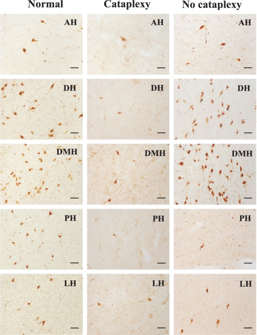
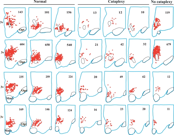
Design: We examined postmortem brain samples, including the hypothalamus of 5 narcolepsy with cataplexy patients; one narcolepsy without cataplexy patient whose complete hypothalamus was available (patient 1); one narcolepsy without cataplexy patient with anterior hypothalamus available (patient 2); and 6 normal brains. The hypothalamic tissue was immunostained for Hcrt-1, melanin-concentrating hormone (MCH), and glial fibrillary acidic protein (GFAP).

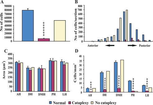
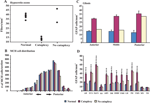
Measurements and results: Neither of the narcolepsy without cataplexy patients had a loss of Hcrt axons in the anterior hypothalamus. The narcolepsy without cataplexy patient whose entire brain was available for study had an overall loss of 33% of hypocretin cells compared to normals, with maximal cell loss in the posterior hypothalamus. We found elevated levels of gliosis with GFAP staining, with levels increased in the posterior hypothalamic nucleus by (295%), paraventricular (211%), periventricular (123%), arcuate (126%), and lateral (72%) hypothalamic nuclei, but not in the anterior, dorsomedial, or dorsal hypothalamus. There was no reduction in the number of MCH neurons in either patient.

Conclusions: Narcolepsy without cataplexy can be caused by a partial loss of hypocretin cells.


Figures

Figure 1
Figure 1
Hypocretin cell loss in narcolepsy with and without cataplexy. A, The total number of hypocretin cells was estimated by stereology in normals and narcoleptics with and without cataplexy (patient 1). Ninety two percent of hypocretin cells were lost in narcolepsy with cataplexy. The patient having narcolepsy without cataplexy (patient 1) had a 33% overall loss in hypocretin cells. B, Hcrt cell distribution in normal, narcolepsy with cataplexy and narcolepsy without cataplexy (patient 1). Hcrt cells were mapped in individual sections from anterior to posterior hypothalamus with 1200 μm inter-section interval. C, The size of the hypocretin cells was estimated by the nucleator method. Surviving hypocretin cells in narcolepsy with and without cataplexy did not differ in size from those in normal brains. D, Hypocretin cell density in the hypothalamic nuclei. In patient 1, who had narcolepsy without cataplexy cell loss was maximal in the posterior hypothalamus, with only 5% of the normal number of these cells present in this region. AH, anterior hypothalamus; DH, dorsal hypothalamus; DMH, dorsomedial hypothalamus; LH, lateral hypothalamus; PH, posterior hypothalamus. (Significance compared to normal, ****P < 0.0001, ***** P < 0.00001).
Figure 2
Figure 2
Hypocretin cells in the hypothalamic nuclei of normals and narcoleptics with and without cataplexy. In normals, hypocretin cell somas are localized in AH, DH, DMH, PH, and LH nuclei. In narcolepsy with cataplexy, cell loss was found in AH, DH, DMH, PH, and LH nuclei, whereas, in narcolepsy without cataplexy (patient 1) cell loss was limited to PH and LH nuclei. (Abbreviations are same as in Figure 1).
Figure 3
Figure 3
Neurolucida mapping of hypocretin cells in 3 normals, 3 narcoleptics with cataplexy, and 1 narcoleptic without cataplexy (patient 1). The cell counts are listed in the upper right hand corner of each section. 3v, third ventricle; Fx, fornix; Mmb, mammillary body; Opt, optic tract.
Figure 4
Figure 4
Hypocretin axon density, MCH distribution and gliosis. A, Hypocretin fiber density fibers/mm2) in the anterior hypothalamus of normals, narcolepsy with cataplexy patients and narcolepsy without cataplexy patients (patients 1 and 2). Hypocretin axon density in the anterior hypothalamus is within the normal range in both of the narcolepsy without cataplexy patients (patients 1 and 2). B, MCH cell distribution in normals, narcolepsy with cataplexy and narcolepsy without cataplexy (patient 1). MCH cells were mapped in individual sections from anterior to posterior hypothalamus with 1200 μm inter-section interval. MCH cell number is normal in narcolepsy with and without cataplexy. C, GFAP density (cells/mm2) in the hypothalamus of normal and narcoleptic brains. The data are grouped into anterior, middle, and posterior regions of the hypothalamus. In narcolepsy without cataplexy (patient 1), increased number of GFAP cells were found only in the posterior hypothalamus. D, GFAP density in different nuclei of the hypothalamus of normal and narcoleptic brains (significance compared to normal, *P < 0.05, **P < 0.01, ***P < 0.001, ****P < 0.0001). AH, anterior hypothalamus; PVN, periventricular nucleus; PAVN, paraventricular nucleus; SO, supraoptic; ARN, arcuate nucleus; DH, dorsal hypothalamus; DMH, dorsomedial hypothalamus; VMH, ventromedial hypothalamus; LH, lateral hypothalamus; PH, posterior hypothalamus; TMN, tuberomammillary nucleus; MN, mammillary nucleus; TH, thalamus.

Similar articles

Cited by

References

    1. Billiard M. Diagnosis of narcolepsy and idiopathic hypersomnia. An update based on the International Classification of Sleep Disorders. Sleep Med Rev. (2nd ed.) 2007;11:377–88. - PubMed
    1. Benca RM. Narcolepsy and excessive daytime sleepiness: diagnostic considerations, epidemiology, and comorbidities. J Clin Psychiatry. 2007;68(Suppl 13):5–8. - PubMed
    1. Longstreth WT, Jr, Koepsell TD, Ton TG, Hendrickson AF, van BG. The epidemiology of narcolepsy. Sleep. 2007;30:13–26. - PubMed
    1. Guilleminault C. Cataplexy. In: Guilleminault C, Dement WC, Passouant P, editors. Narcolepsy. 3rd ed. New York: Spectrum; 1976. pp. 125–43.
    1. Blouin AM, Thannickal TC, Worley PF, Baraban JM, Reti IM, Siegel JM. Narp immunostaining of human hypocretin (orexin) neurons: Loss in narcolepsy. Neurology. 2005;65:1189–92. - PMC - PubMed

Publication types