

Review
Ocular lymphatics: state-of-the-art review
Affiliations
- PMID: 19725271
- PMCID: PMC4725303
Item in Clipboard
Review
Ocular lymphatics: state-of-the-art review
Lymphology.
2009 Jun.
Abstract
Research involving the lymphatic system has experienced an exponential progression during the past decade largely because of advancement of modern technology and discovery of several lymphatic specific molecules. The eye provides an excellent site for lymphatic studies due to its accessible location and the unique feature of tissue heterogeneity--while some tissues are lymphatic-rich, others are lymphatic-free or -inducible. This review provides an update on our current understanding of ocular lymphatics and possible associated eye diseases.
Figures
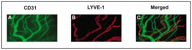
Representative micrographs showing newly developed lymphatics (LYVE-1highCD31low) in the inflamed cornea. Red: LYVE-1 (a lymphatic marker); Green: CD-31 (a panendothelial marker); Merged: yellow. Original magnification: X100.
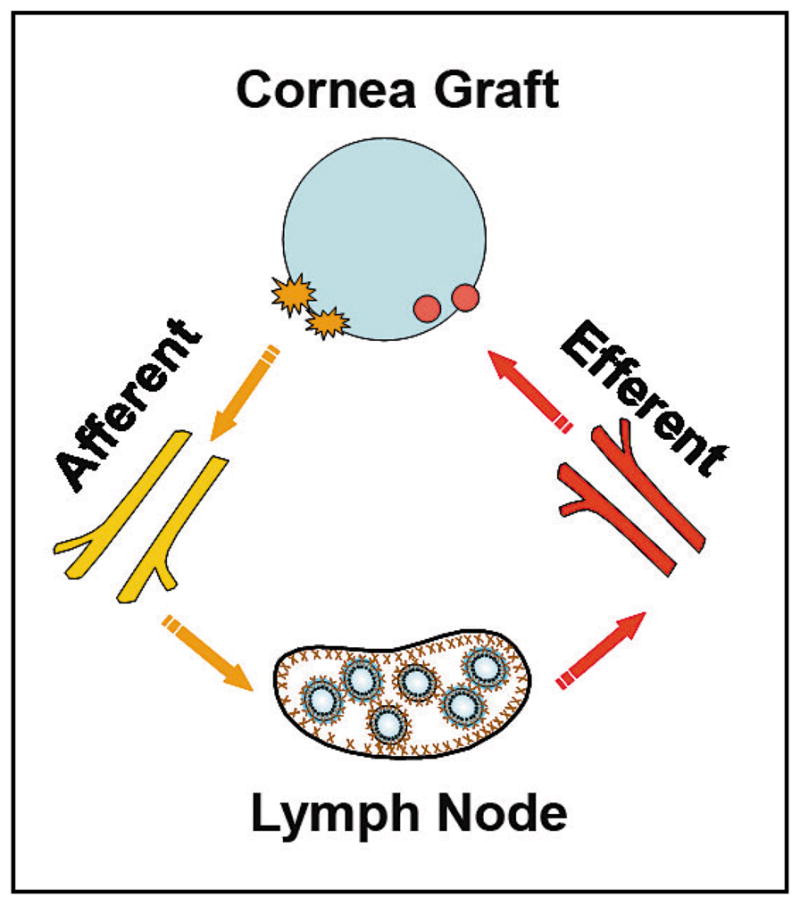
Importance of lymphatic and blood vessels as entry and exit processes of the immune reflex arc involved in corneal transplant rejection. Yellow: lymphatics of the afferent pathway facilitating antigen-presenting cell trafficking; Red: blood vessels of the efferent pathway facilitating T cell infiltration to the graft.
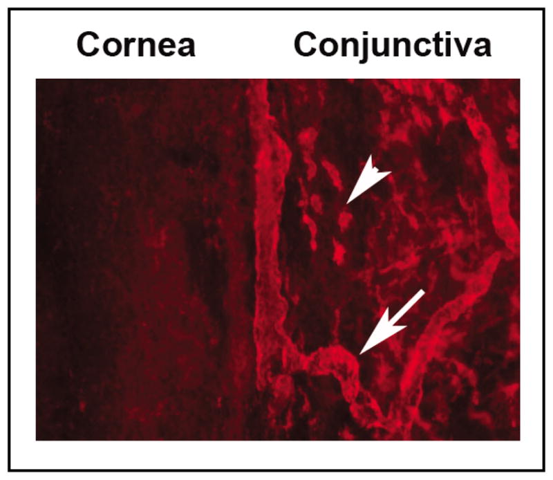
Representative micrographs demonstrating the lymphatic-rich (arrow) conjunctiva and the lymphatic-free cornea. A number of LYVE-1+ cells (arrowhead) also exist in the limbus and conjunctiva. Original magnification: X100.
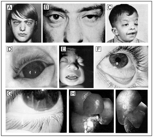
Ocular abnormalities in hereditary lymphedema syndromes. A. Ptosis, conjunctival edema, and medial eyebrow flare in Lymphedema-hypoparathyroidism syndrome. B. Unilateral ptosis in Lymphedema-ptosis syndrome. C. Ptosis and hypertelorism in Noonan syndrome with intestinal lymphangiectasia. D. Ectropion uveae and E. Bilateral subepicanthal folds with facial edema in Persistent mullerian derivatives with intestinal lymphangiectasia. F. Conjunctival edema and chemosis in Mucke syndrome. G. Distichiasis and chemosis in Lymphedema-distichiasis syndrome. H. Punched-out lesions in the retina of siblings with Chorioretinal dysplasia-lymphedema syndrome. Composite reproduced with permission from Northup et. al., Lymphology (87) with original permissions and citations.
References
-
- Aselli G. De lactibus sive lacteis venis. Milan. Mediolani: 1627.
-
- Alitalo K, Tammela T, Petrova TV. Lymphangiogenesis in development and human disease. Nature. 2005;438:946–953. - PubMed
-
- Brown P. Lymphatic system: unlocking the drains. Nature. 2005;436:456–458. - PubMed
-
- Cursiefen C, Chen L, Dana MR, et al. Corneal lymphangiogenesis: Evidence, mechanisms, and implications for corneal transplant immunology. Cornea. 2003;22:273–281. - PubMed
-
- Folkman J, Kaipainen A. Genes tell lymphatics to sprout or not. Nat Immunol. 2004;5:11–12. - PubMed
Publication types
MeSH terms
Grants and funding
LinkOut - more resources
Full Text Sources
Other Literature Sources
Medical
Miscellaneous