. 2009 Dec;88(12):711-7.
doi: 10.1016/j.ejcb.2009.08.001. Epub 2009 Sep 2.

Golgi polarity does not correlate with speed or persistence of freely migrating fibroblasts

Affiliations

Golgi polarity does not correlate with speed or persistence of freely migrating fibroblasts

Andrea C Uetrecht et al. Eur J Cell Biol. 2009 Dec.

Abstract

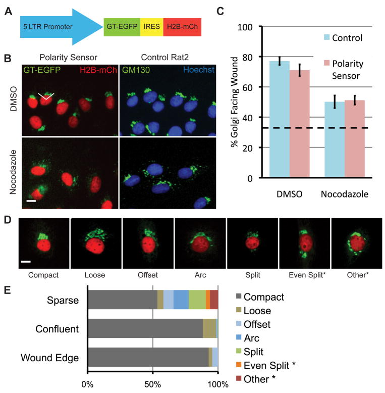
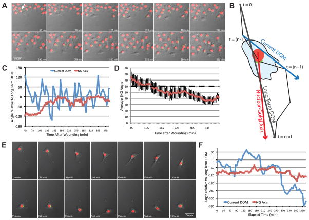
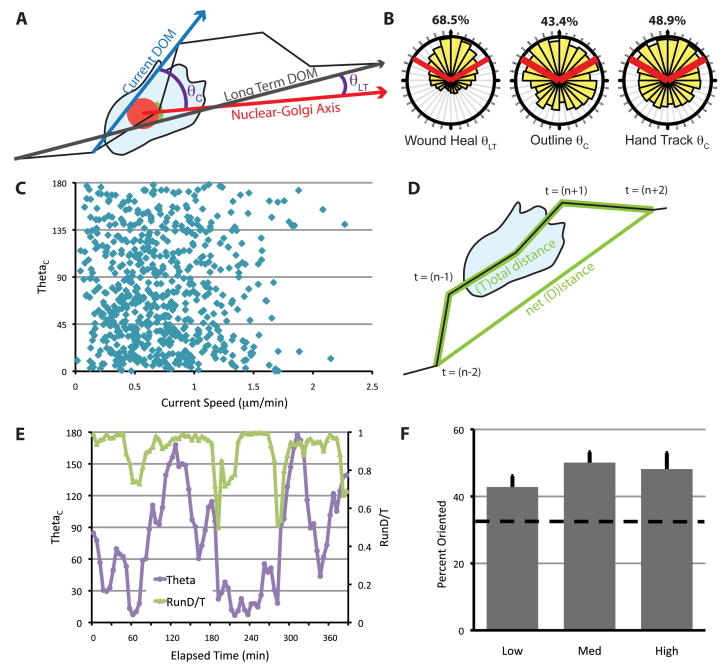
The polarization of the Golgi has long been thought to be important for cell migration. Here we show that Rat2 cells at the edge of an artificial wound repolarize the Golgi relative to the nucleus to face the direction of migration into the wound. However, in the absence of cues from neighboring cells, individual cells do not display Golgi polarity relative to the direction in which they are moving. Instead, the positioning of the Golgi relative to the nucleus remains relatively constant over time and does not reflect changes in the direction of migration. Consistent with this observation, we observe only a slight bias in Golgi positioning to the front of the nucleus, and this bias is not higher during periods of time when the cell is moving in a persistent manner. Taken together, these data suggest that Golgi polarity is not a requirement for cell migration.


Figures

Fig. 1
Fig. 1
Characterization of the polarity sensor. (A) Polarity sensor design. The Golgi is marked with the first 81 amino acids of GT fused to EGFP. The nucleus is marked with H2B fused to mCherry. These sequences flank an internal ribosomal entry sequence (IRES) to allow simultaneous expression from a single promoter. (B) Representative images of cells in the scratch-wound assay treated with either DMSO (control) or nocodazole (0.1 μg/ml). Uninfected Rat2 cells are labeled with anti-GM130 (green) and Hoechst (blue) to mark the Golgi and nucleus, respectively. The wound edge is up, the white lines indicate ±60° facing the wound edge, considered polarized. Scale bar = 10 μm. (C) Quantification of results in (B). Results are from at least 100 cells per treatment from each of 3 independent experiments. Data were analyzed using one-way ANOVA and Tukey’s post-hoc test. Error bars = S.E.M.; dashed line indicates random Golgi positioning. (D) Golgi morphology is heterogeneous. Representative images of different Golgi morphologies observed in sparsely plated cells. Scale bar = 10 μm. (E) Quantification of results in (D). Two categories of Golgi morphology, indicated by asterisks, were excluded from subsequent analyses because the Golgi position could not be determined.
Fig. 2
Fig. 2
The NGA aligns with the direction of migration into the wound but does not align with the direction of migration in freely migrating cells. (A) Representative time-lapse images of Rat2 cells expressing the polarity sensor in the scratch-wound assay. The arrow indicates the cell analyzed in (C). (B) Schematic diagram of axes used for analysis. The cell track is represented by the black line, from t=0 to t=end. These two points define the long-term DOM (turquoise arrow). The current DOM at t=n is defined by the position of the cell centroid at t=(n−1) and t=(n+1) (blue arrow). The NGA is the vector defined by the current nuclear and Golgi positions (red arrow). (C) Current DOM and NGA relative to the long-term DOM over time for the representative cell indicated in (A). (D) The average absolute value of NGA relative to the long-term DOM for multiple cells (n~60) in the wound healing assay over time. Error bars = S.e.M. (E) Time-lapse images of a freely migrating cell expressing the polarity sensor. (F) Current DOM and NGA of the cell in (E) relative to the long-term DOM over time. In this case, DOM was calculated from the hand-outlined track.
Fig. 3
Fig. 3
Nucleus-Golgi polarity is not correlated with the direction of migration in freely migrating cells. (A) Schematic for calculating θ. θLT is used for cells in the scratch wound assay and θC is used for freely migrating cells. The current DOM (and θC) was calculated from cell centroids determined in two ways, as described in the text and Fig. S1. (B) The distribution of θ for cells at the wound edge or in freely migrating cells. Red lines indicate ±60° facing the DOM, and the percentage of data that fall within these boundaries are indicated above. Data were generated from at least three independent experiments with at least 100 cells total for each condition, tracked over at least 5 h. (C) θ as a function of current cell speed. Current cell speed was calculated using tracks from outlined cells over 10 min from (t−1) to (t+1). (D) RunD/T was calculated as a sliding window using cell centroids from time (t−2), (t−1), (t), (t+1) and (t+2). D/T is defined as the net path length (D) divided by the total path length (T). (E) θ and RunD/T for the cell shown in Fig. 2E over time. RunD/T was calculated from the hand-outlined track of this cell. (F) Percentage of times when the Golgi is oriented (θ < 60°), during times when RunD/T is high (≥ 0.9), medium (0.9 > RunD/T > 0.7) or low (< 0.7) in freely migrating cells. RunD/T was calculated from the hand-tracked positions, and instances when cells were deemed too slow for accurate tracking were eliminated (see Fig. S1). The dashed line indicates random Golgi positioning. Error bars = S.E.M. Data were generated from four independent experiments with at least 100 cells total.

References

    1. Bear JE, Svitkina TM, Krause M, Schafer DA, Loureiro JJ, Strasser GA, Maly IV, Chaga OY, Cooper JA, Borisy GG, Gertler FB. Antagonism between Ena/VASP proteins and actin filament capping regulates fibroblast motility. Cell. 2002;109:509–521. - PubMed
    1. Cai L, Marshall TW, Uetrecht AC, Schafer DA, Bear JE. Coronin 1B coordinates Arp2/3 complex and cofilin activities at the leading edge. Cell. 2007;128:915–929. - PMC - PubMed
    1. Danowski BA, Khodjakov A, Wadsworth P. Centrosome behavior in motile HGF-treated PtK2 cells expressing GFP-gamma tubulin. Cell Motil Cytoskeleton. 2001;50:59–68. - PubMed
    1. De Wever O, Westbroek W, Verloes A, Bloemen N, Bracke M, Gespach C, Bruyneel E, Mareel M. Critical role of N-cadherin in myofibroblast invasion and migration in vitro stimulated by colon-cancer-cell-derived TGF-beta or wounding. J Cell Sci. 2004;117:4691–4703. - PubMed
    1. Desai RA, Gao L, Raghavan S, Liu WF, Chen CS. Cell polarity triggered by cell-cell adhesion via E-cadherin. J Cell Sci. 2009;122:905–911. - PMC - PubMed

Publication types

LinkOut - more resources