. 2009 Nov;83(22):11734-45.
doi: 10.1128/JVI.00578-09. Epub 2009 Sep 2.

Induction of therapeutic antibodies by vaccination against external loops of tumor-associated viral latent membrane protein

Affiliations

Induction of therapeutic antibodies by vaccination against external loops of tumor-associated viral latent membrane protein

Catherine Delbende et al. J Virol. 2009 Nov.

Abstract

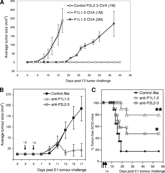
Some human herpesviruses (HHV) are etiological contributors to a wide range of malignant diseases. These HHV express latent membrane proteins (LMPs), which are type III membrane proteins consistently exposed at the cell surface in these malignancies. These LMPs have relatively large cytoplasmic domains but only short extracellular loops connecting transmembrane segments that are accessible at the surface of infected cells, but they do not elicit antibodies in the course of natural infection and tumorigenesis. We report here that conformational peptides mimicking two adjacent loops of the Epstein-Barr virus (EBV) LMP1 (2LS peptides) induce high-affinity antibodies with remarkable antitumor activities in mice. In active immunization experiments, LMP1-targeting 2LS vaccine conferred tumor protection in BALB/c mice. Moreover, this tumor protection is dependent upon a humoral anti-2LS immune response as demonstrated in DO11.10 (TCR-OVA) mice challenged with LMP1-expressing tumor and in SCID mice xenografted with human EBV-positive lymphoma cells. These data provide a proof of concept for 2LS immunization against short external loops of viral LMPs. This approach might possibly be extended to other infectious agents expressing type III membrane proteins.


Figures

FIG. 1.
FIG. 1.
Design of 2LS conformational epitopes and characteristics of P1L1-2 Abs. (A) Topology prediction for LMP1, indicating 2LS peptide construction strategy and the region targeted by the Abs. The Cys residue located centrally in each peptide is used to combine residues from two adjacent external loops (circled letters) and to branch the resulting construct to KLH or OVA. (B) Determination of P1L1-2 Ab specificity by competitive ELISA at various concentrations (ao, 10−10 to 10−6 M) of P1L1-2, P1L2-3, and P1L1-3 peptides. Sera were collected from BALB/c mice (five per group) a week after each P1L1-2 vaccine dose and tested at a 1/150 dilution for binding to peptides. At equilibrium, the mixture was analyzed by direct ELISA with the three peptides as solid-phase antigen for only the third vaccine dose. (C) The displacement curves obtained in panel B by fitting A/Ao to ao were linearized by A/(AoA) to 1/ao transformation. Ab affinity is proportional to the slopes of initial curve segments and increases with the number of doses of vaccines in a representative P1L1-2-vaccinated mouse group. The slope for each curve, corresponding to Ab affinity for collected sera, is indicated. Results are representative of four independent experiments.
FIG. 2.
FIG. 2.
P1L1-2 Abs recognizes native LMP1 and presents functional in vitro activities. (A) To test binding of P1L1-2 Abs to native LMP1, HEK293 cells were stably transfected with pREP4-LMP1 vector (pLMP1-HEK). As revealed by staining, P1L1-2 Abs reacted with all pLMP1-HEK cells but not with control vector-transfected HEK cells (magnification, ×400). (B) To confirm specific binding of P1L1-2 Abs to wild-type LMP1 sequence, cell lysates of pSV-HA-LMP1-transfected HEK293 cells (LMP1-HEK) or vector-HEK293 cells (vector-HEK) were subjected to immunoprecipitation (IP) with P1L1-2 Abs and the commercial CS1-4 Ab, a monoclonal Ab directed to a cytoplasmic cryptic domain of LMP1 or NMS. Immunoprecipitates and total cell extracts of LMP1-HEK (Ptotal) were tested for the presence of LMP1 by immunoblotting using S12, a monoclonal anti-LMP1 with characteristics similar to those of CS1-4. No immunoprecipitate was observed with either extracts from vector-HEK, used as negative control, or NMS. (C) Binding of P1L1-2 Abs to live cell lines resulting from EBV-driven (LCLs and E1 cells) or pREP4-LMP1-induced (LMP1-HEK) LMP1 expression was monitored by cytofluorimetric analysis. Binding of P1L1-2 abs was shown on LCL, E1, and pLMP1-HEK cell lines, which expressed natural or transfected endogenous LMP1, but not on DG75, an EBV-negative Burkitt's lymphoma line, or on a pREP4-transfected HEK cell line. (D) Growth inhibition and cell aggregation of LCL cells incubated with anti-P1L1-2 Abs for 4 days. Cell aggregation was not observed with control mouse serum (upper), whereas metabolic arrest and cell aggregation were visualized without magnification for cells treated with anti-P1L1-2 Abs (lower). (E) LCL, E1, and DG75 cell viabilities were assessed after 4 days of culture with P1L1-2 Abs from P1L1-2-KLH-immunized mice at the indicated dilutions in the presence of a large excess of NMS (1/60) with active complement as described in the text. Data are means ± standard errors of the means from three independent experiments. *, significantly different from control serum (*, P < 0.5; **, P < 0.001 [Student's t test]).
FIG. 3.
FIG. 3.
P1L1-2 vaccine induces Abs targeting cell surface LMP1 and protection of BALB/c mice from tumors. (A) LMP1 expression was assessed in LCLs, E1 cells, and the F2 cell line, obtained by selection of pREP4-LMP1-transfected Sp2/O cells. Equimolar amounts of cell lysates were tested for LMP1 expression by immunoblot analysis using S12, a monoclonal Ab reacting against a cytoplasmic tail of LMP1. The pSV-HA-LMP1-transfected HEK293 cells (HEKLMP1) overexpressing LMP1 were used as positive control. DG75 is an EBV-negative Burkitt's lymphoma line, and Sp2/O and HEK were transfected with the vectors alone. (B) Extracellular binding of P1L1-2 Abs to LMP1 was revealed by F2 cell staining. Sp2/O cells transfected with vector alone were used as a control (magnification, ×400). (C) To demonstrate that LMP1 was the target of P1L1-2 Abs at the surface of stained cells, live F2 cells and LCLs were incubated with P1L1-2 Abs, washed, and subjected to lysis, and bound Abs were immunoprecipitated (IP) as described in the text. LMP1 molecules from cell surface were detected in immunoprecipitates by immunoblot analysis using S12. HEKLMP1 cell extracts (Ptotal) were used to locate the 63-kDa LMP1 band in the immunoblot. (D) BALB/c mice (six per group) were immunized s.c. three and four times, 2 weeks apart, with the P2L2-3-KLH or P1L1-2-KLH vaccine. Two weeks after the final immunization, mice were challenged with 6 × 10⁶ SP2/O or F2 tumor cells s.c. and followed for surface tumor growth with a caliper (mm2). Mice were euthanized when tumors reached a size of 200 mm2 or become ulcerated. If individual mice within a group were euthanized, the final measurement was carried over to subsequent time points. All control mice immunized with P2L2-3-KLH or P1L1-2-KLH vaccine were euthanized, respectively, by day 19 or 33. Mouse groups treated with the high (three boosts) or low (two boosts) P1L1-2 vaccination protocol were completely or partially protected from F2 tumor development. All mice which did not develop tumor by day 30 remained tumor free beyond 150 days. Error bars represent standard errors of the means. Results are representative of at least three independent experiments.
FIG. 4.
FIG. 4.
P1L1-2 vaccine can induce a partial curative response in BALB/c mice. Groups of six BALB/c mice were challenged with 6 × 10⁶ LMP1-expressing F2 tumor cells and immunized with an irrelevant P2L2-3-KLH control vaccine or the anti-LMP1 P1L1-2-KLH vaccine. Mice were followed for surface tumor growth with a caliper (mm2) and euthanized when tumors reached a size of 200 mm2 or become ulcerated. If individual mice within a group were euthanized, the final measurement was carried over to subsequent time points. (A) Therapeutic vaccination did not protect against tumor challenge. During F2 tumor challenge, two groups of six mice were injected three times with P2L2-3-KLH or P1L1-2-KLH vaccine as indicated by the arrows. All control and anti-LMP1-treated mice were euthanized by day 23. (B) Therapeutic vaccination of preimmunized mice partially protects against tumor challenge. Two groups of six mice were injected with P2L2-3-KLH or P1L1-2-KLH vaccine and received their second boost 2 weeks before F2 tumor challenge (low-vaccination protocol). At day 23, mice received a therapeutic P1L1-2-KLH vaccine dose (arrows, +1). (C) Individual tumor growth in the P1L1-2-KLH-vaccinated mice represented in panel B. Two mice developed low or high tumor progression. The mouse with low tumor progression regressed (mouse 1), and that with high tumor progression had to be euthanized by day 35 (mouse 2). The mice which controlled tumor (mice 1 and 3 to 6) remained tumor free beyond 150 days. (D) Isotype profiles and serum levels of P1L1-2 Abs in the experiment represented in panels B and C. Circulating IgG isotypes were determined at day 15 before tumor appearance for each mouse (mice 1 to 6) and at day 30 for mice with tumor regression (mouse 1) or tumor development (mouse 2). Data are expressed as mean ± standard error of the mean for six mice per group. Results are representative of three independent experiments.
FIG. 5.
FIG. 5.
P1L1-2 vaccine induces P1L1-2 Abs that protect from tumors in TCR-OVA mice and SCID mice xenografted with human E1 cells. (A) Groups of six TCR-OVA mice received s.c., 2 week apart, two doses of P1L1-2-OVA or P2L2-3-OVA vaccine. Five days after the final immunization, mice were challenged with 1 or 3 million F2 tumor cells (1 M, 3 M) s.c. and were followed for surface tumor growth with a caliper (mm2). P1L1-2-immunized TCR-OVA T-cell-deficient mice were protected from tumor challenge compared to P2L2-3-immunized controls (P < 0.0001). A threefold increase in tumor cell number was necessary to observe tumor development in P1L1-2-immunized TCR-OVA mice. (B and C) Mean tumor size evolution (B) and survival of SCID mice (C) (10/group) challenged with EBV-infected human E1 cells and subsequent i.p. injections by passive Ab transfers with P1L1-2, P2L2-3, or KLH control immune sera. SCID mice were treated at days 3 and 5 after tumor challenge as indicated by the arrows, at a moment where tumors became palpable. Only 20% of control animals survived at day 60 (not requiring euthanasia), whereas treatment with P2L2-3 Ab led to 50% survival, and mice treated with P1L1-2 Ab survived up to 80%. (*, P2L2-3 Abs versus control [P < 0.5]; **, P1L1-2 Abs versus control [P < 0.001]). Error bars represent standard errors of the means. Results are representative of at least three independent experiments.

Similar articles

Cited by

References

    1. Beisser, P. S., L. Laurent, J. L. Virelizier, and S. Michelson. 2001. Human cytomegalovirus chemokine receptor gene US28 is transcribed in latently infected THP-1 monocytes. J. Virol. 75:5949-5957. - PMC - PubMed
    1. Bernstein, D. I. 2001. Potential for immunotherapy in the treatment of herpesvirus infections. Herpes 8:8-11. - PubMed
    1. Brinkmann, M. M., and T. F. Schulz. 2006. Regulation of intracellular signalling by the terminal membrane proteins of members of the Gammaherpesvirinae. J. Gen. Virol. 87:1047-1074. - PubMed
    1. Burton, D. R. 2002. Antibodies, viruses and vaccines. Nat. Rev. 2:706-713. - PubMed
    1. Carter, P. J. 2006. Potent antibody therapeutics by design. Nat. Rev. Immunol. 6:343-357. - PubMed

Substances

LinkOut - more resources