. 2009 Nov;83(22):11447-55.
doi: 10.1128/JVI.00748-09. Epub 2009 Sep 2.

Receptor binding and low pH coactivate oncogenic retrovirus envelope-mediated fusion

Affiliations

Receptor binding and low pH coactivate oncogenic retrovirus envelope-mediated fusion

Marceline Côté et al. J Virol. 2009 Nov.

Abstract

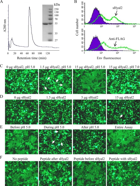
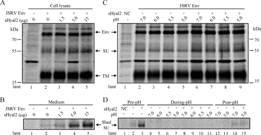
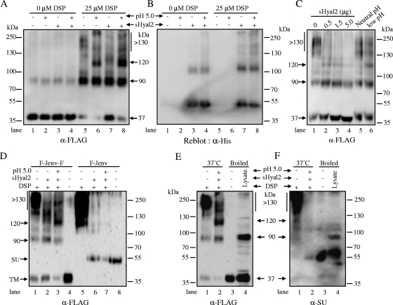
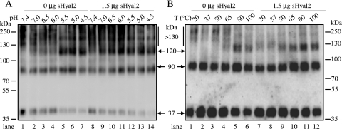
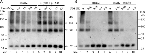
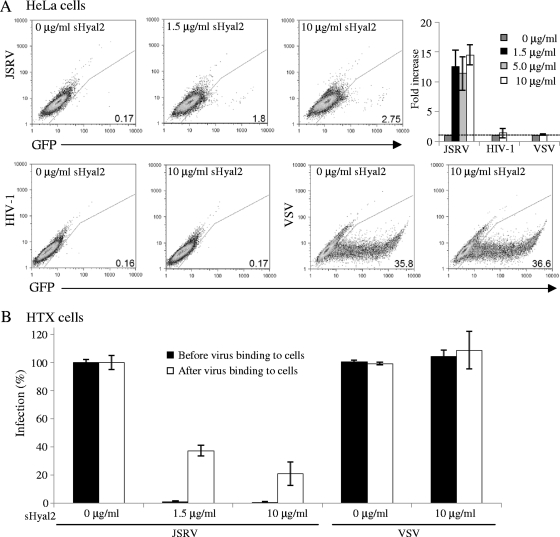
Fusion of enveloped viruses with host cells is triggered by either receptor binding or low pH but rarely requires both except for avian sarcoma leukosis virus (ASLV). We recently reported that membrane fusion mediated by an oncogenic Jaagsiekte sheep retrovirus (JSRV) envelope (Env) requires an acidic pH, yet receptor overexpression is required for this process to occur. Here we show that a soluble form of the JSRV receptor, sHyal2, promoted JSRV Env-mediated fusion at a low pH in normally fusion-negative cells and that this effect was blocked by a synthetic peptide analogous to the C-terminal heptad repeat of JSRV Env. In contrast to the receptor of ASLV, sHyal2 induced pronounced shedding of the JSRV surface subunit, as well as unstable conformational rearrangement of its transmembrane (TM) subunit, yet full activation of JSRV Env fusogenicity, associated with strong TM oligomerization, required both sHyal2 and low pH. Consistently, sHyal2 enabled transduction of nonpermissive cells by JSRV Env pseudovirions, with low efficiency, but substantially blocked viral entry into permissive cells at both binding and postbinding steps, indicating that sHyal2 prematurely activates JSRV Env-mediated fusion. Altogether, our study supports a model that receptor priming promotes fusion activation of JSRV Env at a low pH, and that the underlying mechanism is likely to be different from that of ASLV. Thus, JSRV may provide a useful alternate model for the better understanding of virus fusion and cell entry.


Figures

FIG. 1.
FIG. 1.
sHyal2 mediates syncytium formation by JSRV Env in 293 cells at low pH, the effect of which is inhibited by a synthetic C-HR2 peptide. (A) Size exclusion chromatography and SDS-PAGE of sHyal2. sHyal2 (∼50 kDa) was purified by fast protein liquid chromatography, and the purified sHyal2 was analyzed by SDS-PAGE and Coomassie blue staining. (B) sHyal2 specifically binds to 293 cells expressing a FLAG-tagged JSRV Env (top); expression of the latter on the cell surface was confirmed by an anti-FLAG antibody (bottom). (C) Syncytium formation by JSRV Env in 293 cells (expressing GFP) is induced only by sHyal2 plus pH 5.0 (central panels), but not by either one alone (left and right panels). (D) A JSRV Env chimera, containing the ENTV SU and with reduced binding to Hyal2, requires an increased concentration of sHyal2 for fusion (see the text for details). (E) sHyal2 acts before but not after the pH 5.0 pulse for fusion induction. These experiments employed 5 μg sHyal2. (F) The JSRV C-HR2 peptide acts after sHyal2 incubation and before a low-pH pulse to inhibit fusion. The assays employed 5 μg sHyal2 and 30 μg C-HR2 peptide, respectively. Arrows are placed to indicate the fused cells only when fusion is not obvious.
FIG. 2.
FIG. 2.
sHyal2, but not low pH, promotes shedding of JSRV SU. (A and B) 293 cells expressing JSRV Env were metabolically labeled for 1 h and then chased for 3 h before the addition of the indicated amounts of sHyal2. Cells were chased for an additional 3 h before being lysed. Cell lysates and culture media were harvested and immunoprecipitated using anti-FLAG beads. Samples were resolved by SDS-PAGE and analyzed by autoradiography. (A) Env expression and processing in cell lysates. (B) JSRV SU shedding into the culture medium. (C and D) Metabolic labeling was performed as described for panels A and B, except that cells were incubated in the absence or presence of 5 μg sHyal2. The culture medium was harvested (pre-pH), and the cells were treated with the indicated pH buffers for 5 min at 37°C. The pH buffers were harvested and neutralized (during pH), and the cells were cultured for an additional 1 h. The culture medium was harvested (post-pH), and the cells were lysed. The cell lysates (C) and harvested supernatants or neutralized pH buffers (D) were immunoprecipitated using anti-FLAG beads and were analyzed by autoradiography. Env, JSRV Env precursor; NC, parental 293 cells not expressing JSRV Env. The strong band appearing in the parental 293 cells (panels A and C, lanes 1), and sometimes in the Env-expressing 293 cells, is likely a cellular protein that was nonspecifically pulled down by anti-FLAG beads.
FIG. 3.
FIG. 3.
JSRV TM oligomerization induced by sHyal2 and low pH. (A) Purified JSRV Env pseudovirions were incubated with or without 1.5 μg of sHyal2, followed by treatment with a pH 7.4 or pH 5.0 buffer. Samples were either cross-linked by 25 μM DSP or left untreated, and TM oligomerization was analyzed by Western blotting using an anti-FLAG (α-FLAG) antibody. (B) The membrane was then stripped and reblotted using an anti-His (α-His) tag antibody to detect sHyal2. (C) Effect of sHyal2 on TM oligomerization. Purified virions were incubated with the indicated amounts of sHyal2 or were treated with a neutral or a low pH, which served as negative and positive controls, respectively. Samples were cross-linked and analyzed by Western blotting. (D) Purified JSRV pseudovirions bearing F-Jenv-F or F-Jenv were either treated with 1.5 μg sHyal2, alone or in combination with pH 5.0, or left untreated. Samples were either cross-linked by DSP (lanes 1 to 3 and 5 to 7) or boiled without cross-linking (lanes 4 and 8). Note that fivefold more virus was used for F-Jenv than for F-Jenv-F, in order to enhance the detection of SU. (E and F) Purified pseudovirions bearing F-Jenv-F were either left untreated or treated with 1.5 μg sHyal2 plus pH 5.0; then they were cross-linked with DSP. The cross-linked virus samples (indicated as “37°C”) and boiled virions and cell lysates (“Boiled”) were resolved by SDS-PAGE and analyzed by Western blotting using an anti-FLAG antibody (E) or an anti-JSRV SU (α-SU) antibody (F).
FIG. 4.
FIG. 4.
sHyal2 modulates the thresholds of low pH and temperature required for JSRV TM oligomerization. JSRV pseudovirions, preincubated with 1.5 μg sHyal2 or an equal volume of PBS, were treated with the indicated pH conditions (A) or temperatures (B) for 5 min, followed by cross-linking with DSP, and were analyzed by Western blotting using an anti-FLAG antibody.
FIG. 5.
FIG. 5.
Stability of TM oligomers induced by sHyal2 and low pH. JSRV Env pseudovirions, preincubated either with 1.5 μg sHyal2 alone or with 1.5 μg sHyal2 plus pH 5.0, were subjected to the indicated concentrations of urea (A) or SDS (B) for 5 min at 37°C before being cross-linked by DSP and analyzed by Western blotting.
FIG. 6.
FIG. 6.
sHyal2 enables transduction of nonpermissive cells by JSRV Env pseudovirions (A) but blocks their entry into permissive cells (B). (A) HeLa cells were bound by JSRV Env, HIV-1 Env, or VSV-G pseudovirions encoding GFP by spinoculation, followed by incubation with the indicated amounts of sHyal2. The transduction efficiency was analyzed by flow cytometry 48 h postinfection. The titers of JSRV pseudovirions with the presence of 0, 1.5, and 10 μg of sHyal2 in HeLa cells are 34, 360, and 550 GFP+ cells/ml, respectively. The titers of HIV-1 Env and VSV-G pseudotypes in HeLa cells are ∼35 and 3.6 × 10⁶ GFP+ cells/ml, respectively. Note that the low titer of HIV-1 Env pseudovirions in HeLa cells was not due to the viral stock preparation or infection, because the titer in HeLa-TZM-bl cells expressing CD4 and CCR5 was 5 × 10⁴ GFP+ cells/ml. Representative dot plots are shown, and the percentages of GFP+ cells are given at the lower right. The changes (n-fold) in transduction efficiency in four independent experiments were averaged and plotted. (B) HTX cells either were infected with JSRV Env or VSV-G pseudovirions that had been preincubated (before binding) with the indicated amounts of sHyal2 or were first bound with pseudovirions and then incubated with the indicated amounts of sHyal2 (after binding); see details in Materials and Methods. In both cases, unbound sHyal2 and virions were removed from cells by washing, and viral infectivity was assessed by flow cytometry 48 h postinfection.

References

    1. Bertrand, P., M. Côté, Y.-M. Zheng, L. M. Albritton, and S.-L. Liu. 2008. Jaagsiekte sheep retrovirus utilizes a pH-dependent endocytosis pathway for entry. J. Virol. 82:2555-2559. - PMC - PubMed
    1. Brindley, M. A., L. Hughes, A. Ruiz, P. B. McCray, Jr., A. Sanchez, D. A. Sanders, and W. Maury. 2007. Ebola virus glycoprotein 1: identification of residues important for binding and postbinding events. J. Virol. 81:7702-7709. - PMC - PubMed
    1. Brindley, M. A., and W. Maury. 2005. Endocytosis and a low-pH step are required for productive entry of equine infectious anemia virus. J. Virol. 79:14482-14488. - PMC - PubMed
    1. Caporale, M., C. Cousens, P. Centorame, C. Pinoni, M. De las Heras, and M. Palmarini. 2006. Expression of the Jaagsiekte sheep retrovirus envelope glycoprotein is sufficient to induce lung tumors in sheep. J. Virol. 80:8030-8037. - PMC - PubMed
    1. Carr, C. M., C. Chaudhry, and P. S. Kim. 1997. Influenza hemagglutinin is spring-loaded by a metastable native conformation. Proc. Natl. Acad. Sci. USA 94:14306-14313. - PMC - PubMed

Publication types

LinkOut - more resources