Comparative Study
. 2009 Sep 2;29(35):11043-54.
doi: 10.1523/JNEUROSCI.1924-09.2009.

Neurofilaments bind tubulin and modulate its polymerization

Affiliations
Comparative Study

Neurofilaments bind tubulin and modulate its polymerization

Arnaud Bocquet et al. J Neurosci. .

Abstract

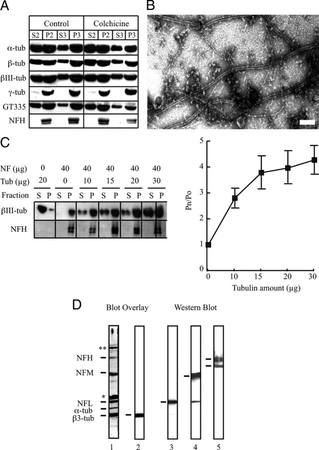
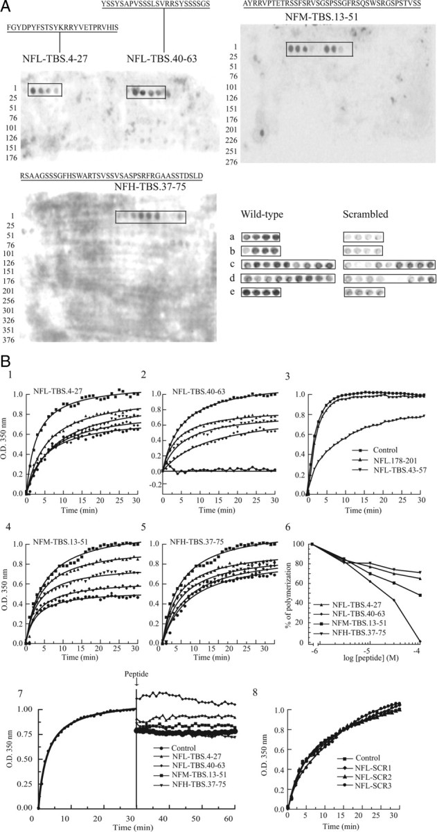
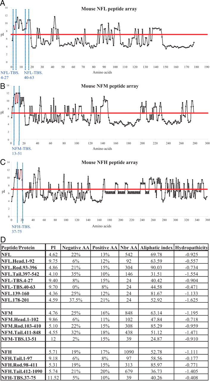
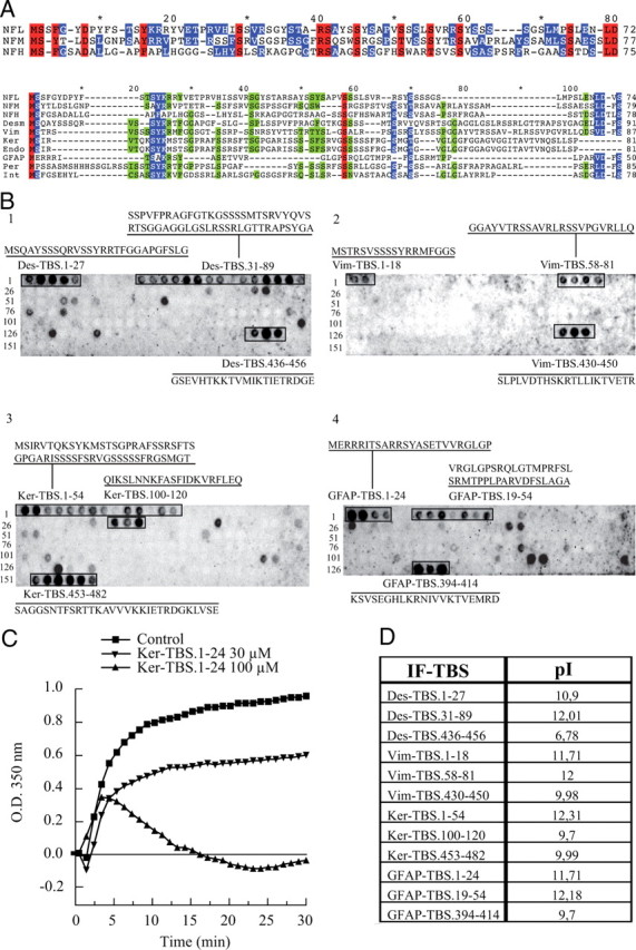
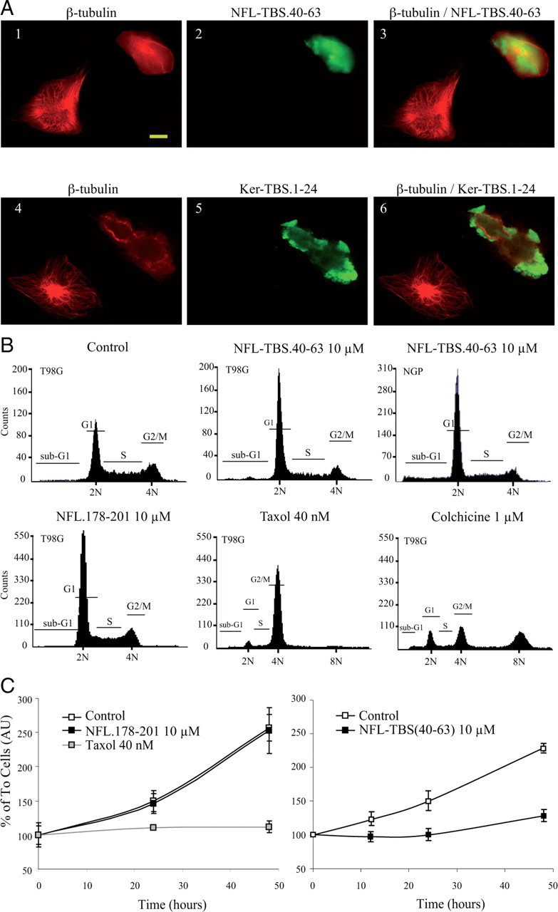
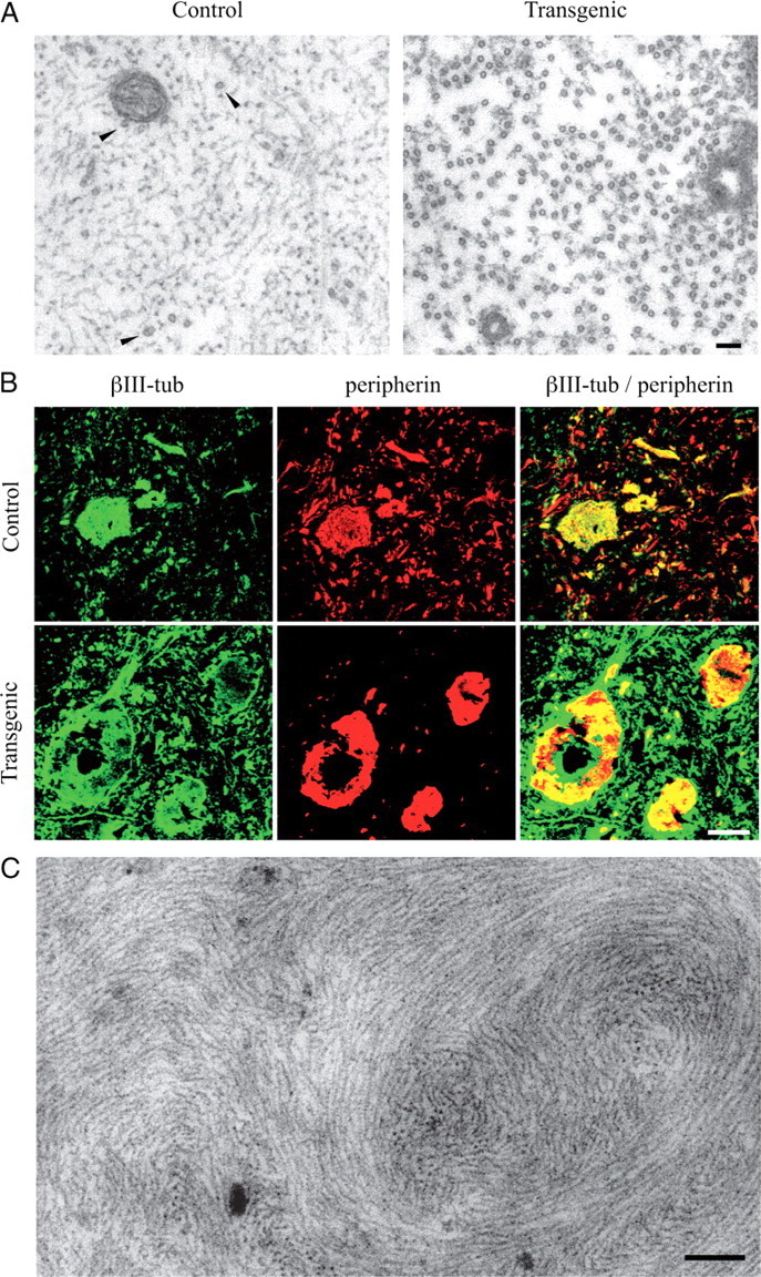
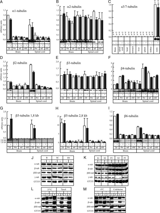
Neurofilaments assemble from three intermediate-filament proteins, contribute to the radial growth of axons, and are exceptionally stable. Microtubules are dynamic structures that assemble from tubulin dimers to support intracellular transport of molecules and organelles. We show here that neurofilaments, and other intermediate-filament proteins, contain motifs in their N-terminal domains that bind unassembled tubulin. Peptides containing such motifs inhibit the in vitro polymerization of microtubules and can be taken up by cultured cells in which they disrupt microtubules leading to altered cell shapes and an arrest of division. In transgenic mice in which neurofilaments are withheld from the axonal compartment, axonal tubulin accumulation is normal but microtubules assemble in excessive numbers. These observations suggest a model in which axonal neurofilaments modulate local microtubule assembly. This capacity also suggests novel mechanisms through which inherited or acquired disruptions in intermediate filaments might contribute to pathogenesis in multiple conditions.


Figures

Figure 1.
Figure 1.
Tubulin coisolates with NFs and is a binding partner of the three NF subunits. A, Tubulin was detected in each fraction of a classical preparation of brain NFs using specific antibodies against α-, β-, βIII-, and γ-tubulin or polyglutamylated epitopes of tubulin (GT335). To prevent MT assembly, all procedures were conducted at 4°C or in the presence of colchicine. B, When the third pellet (P3) from such a NF purification, performed at 4°C but without colchicine, was examined at the electron microscope, no MTs were observed (data not shown), but they were abundant when such preparations are incubated at 37°C in the presence of 1 mm GTP. Scale bar, 100 nm. C, NFs (40 μg) were incubated for 30 min alone (Po) or with increasing quantities of purified tubulin (Pn) and centrifuged, and each pellet and supernatant were analyzed for tubulin content. Tubulin present in the NF pellet increases as an increased amount of tubulin is added. Error bars indicate SEM. D, NFs present in the third pellet (P3) were separated on a 7.5% SDS-PAGE, transferred to a nitrocellulose membrane, and incubated with phosphocellulose-purified tubulin (3 mg/ml). The membrane was incubated with anti-βIII-tubulin and processed like a normal Western blot. The blot overlay (lane 1) revealed that tubulin interacted with bands migrating at the same position as NFL, NFM, and the phosphorylated isoform of NFH, together with α- and β-tubulin, as well as *synapsin and **MAPs. Without the addition of purified tubulin, only the band corresponding to βIII-tubulin was revealed (lane 2). The same membrane was stripped to remove tubulin and anti-βIII-tubulin antibodies, and reprobed sequentially with anti-NFL (lane 3), anti-NFM (lane 4), and anti-NFH (lane 5) antibodies. The anti-NFH antibody recognizes both phosphorylated and dephosphorylated isoforms of NFH. P, Pellet; S, supernatant; tub, tubulin.
Figure 2.
Figure 2.
Tubulin binds to the N-terminal domain of each NF subunit, and peptides corresponding to these TBSs affect MT polymerization. A, Peptide array membranes were incubated with phosphocellulose-purified tubulin (3 mg/ml) overnight at 4°C, and bound tubulin was detected with an anti-βIII-tubulin antibody and a peroxidase-labeled secondary antibody. The NFL subunit contains two 24-aa-long TBSs (NFL-TBS.4-27 and NFL-TBS.40-63), whereas NFM and NFH contain one 39-aa-long TBS (NFM-TBS.13-51 and NFH-TBS.37-75). Bottom, When the sequence of peptides corresponding to the TBS was scrambled (right), their capacity to bind tubulin was typically abolished or greatly diminished. The sequences of peptides are as follows: (a) NFL-TBS (4-27), FGYDPYFSTSYKRRYVETPRVHIS; and NFL-TBS (4-27) scrambled, SDETRHFGVPISKYSYRYPFVYRT; (b) NFL-TBS (40-63), YSSYSAPVSSSLSVRRSYSSSSGS; and NFL-TBS2 (40-63) scrambled, SSASLSYSPSRSVSRSYSGSYSVS; (c) NFM-TBS (13-51), AYRRVPTETRSSFSRVSGSPSSGFRSQSWSRGSPSTVSS; and NFM-TBS (13-51) scrambled, SVWRASYGSRSVSPGTSERTSFSRPSFSRSPRSGVSTQS; (d) NFH-TBS (37-75), RSAAGSSSGFHSWARTSVSSVSASPSRFRGAASSTDSLD; and NFH-TBS (37-75) scrambled, SHSPSTSASRSASWSGSDGRFSADRVATSFSRLGSASAV; (e) Ker-TBS (1-24), MSIRVTQKSYKMSTSGPRAFSSRS; and Ker-TBS1 (1-24) scrambled, QTGAKSISFMRPSVSYRSKTMSRS. B1–B5, Peptides corresponding to the TBS of each NF subunit were added at different concentrations to unpolymerized MTs (S3 fraction from an MT preparation), and their effect on MT assembly was evaluated. ●, 100 μm; ♦, 30 μm; ▼, 10 μm; ▲, 3 μm; or ■, without peptide. B6, The percentage of the MT assembly inhibition after 30 min was plotted against peptide concentration. B7, When peptides were added to fully polymerized MTs (arrow), no depolymerization occurred within 30 min. B8, When the sequence of NFL-TBS.40-63 peptide was scrambled, it lost its capacity to affect MT assembly. The sequences of NFL.SCR1, NFL.SCR2, and NFL.SCR3 are presented in Table 2.
Figure 3.
Figure 3.
Calculation of the pI (isoelectric point) for each peptide spotted on the peptide arrays. A–C, The pI value was calculated for each 15-aa-long peptide present on the peptide array corresponding to NFL (A), NFM (B), and NFH (C). The graphs show that the pI of the TBS is slightly higher when compared with the rest of the sequence. However, some peptides have high pI values, but they do not bind tubulin. D, Several biochemical characteristics of each NF subunit and their intramolecular domains are summarized. Interestingly, the pI value of the “head” domain of each NF subunit is higher compared with the rest of the sequence. Calculations were performed online using “ProtParam tool” available at www.expasy.org.
Figure 4.
Figure 4.
Sequence analysis of the tubulin-binding sites and their presence in other intermediate-filament proteins. A, Sequences of the three mouse NF subunits were aligned together, or with several intermediate-filament sequences, according to their homology by the ClustalW program. Alignment of NF subunits with the other N-terminal domains of intermediate filaments shows several conserved amino acids (red, 100%; blue, 80%; and green, 60% homology). B, Using the same experimental approach as described for NF subunits (compare Fig. 2A), the peptide arrays corresponding to the sequence of several intermediate filaments (desmin, vimentin, keratin, and GFAP) were tested for the possible presence of tubulin-binding sites. For all the intermediate filaments tested, a tubulin-binding site (alignment of 2 or more spots) was located in the N-terminal domain. C, The effect of Ker.TBS.1-24 was tested on MT polymerization, as previously described in Figure 2B, and demonstrates a typical inhibition of MT polymerization. D, Summary of the pI values for each intermediate-filament TBS as calculated online.
Figure 5.
Figure 5.
Effect of NFL-TBS.40-63 and Ker-TBS.1-24 peptides on the MT cytoskeleton and cell division. A, T98G cells were grown in the presence of biotinylated NFL-TBS.40-63 peptide (10 μm) for 6 h (A1–A3). MTs were detected using an anti-tubulin antibody (red), and NFL-TBS.40-63 was detected using Alexa-labeled avidin (green). Cells containing the peptide lack a typical MT network and have a round shape. Similar results were observed using biotinylated Ker-TBS.1-24 (A4–A6). B, Flow cytometric analysis of untreated and NFL-TBS.40-63-treated cells. T98G or NGP cells (third panel) treated with NFL-TBS.40-63 peptides (10 μm) during 48 h showed a typical G1 phase arrest. In the presence of NFL.178-201, no major effect was observed. Taxol and colchicine were used as controls. C, T98G cells were treated with NFL-TBS.40-63, NFL.178-201, or Taxol. Proliferation of NFL-TBS.40-63 peptide-treated cells (10 μm for 48 h) is reduced compared with cells grown in normal media. No effect was observed for NFL.178-201. Error bars indicate SEM.
Figure 6.
Figure 6.
Analysis of the cytoskeleton in neurons of control and NFHLacZ transgenic mice. A, Typical axons in control samples are filled with a densely packed NF cytoskeleton and sparse MTs, whereas in transgenic samples they lack NFs and are filled with MTs. Scale bar, 100 nm. B, Confocal immunofluorescence analysis of spinal cords from control and NFHLacZ transgenic mice with antibodies recognizing βIII-tubulin and peripherin. In NFHLacZ transgenic samples, intermediate-filament labeling is restricted to cell body aggregates, and these aggregates also label with anti-tubulin antibodies. Scale bar, 12.5 μm. C, Electron micrograph of a typical NF aggregate present in a motor neuron cell body in NFHLacZ mice. Despite the presence of tubulin epitopes in such aggregates, no MT profile is detectable. Scale bar, 200 nm.
Figure 7.
Figure 7.
The amount of α- and β-tubulin mRNA and protein is similar between transgenic (+) and control (−) samples. A, During aging, a similar 10-fold decrease of mouse α-tubulin isoform 1 (Mα1) tubulin mRNA occurred both in brain and spinal cord of control and transgenic mice (2d, 2 d; 4w, 4 weeks; ad, adult; old, >24-month-old animals). Mean ± SE is shown. B, Mα2 mRNA remained constant during postnatal development of NFHLacZ mice. C, Mα3/7 tubulin mRNA was detected only in testis. D, Mouse β-tubulin isoform 2 (Mβ2) tubulin mRNA decreased 3-fold in brain and 10-fold in spinal cord in both control and transgenic samples. E, Mβ3 tubulin accumulated to similar levels in both normal and transgenic mice. F, Mβ4 tubulin mRNA showed a two-phase profile in developing brain and spinal cord samples. First, Mβ4 mRNA levels increased in brain threefold between 2 d and 4 weeks, and fivefold in spinal cord. Thereafter, Mβ4 tubulin mRNA decreased by a factor of 2 both in brain and spinal cord. G, H, Two Mβ5 transcripts exhibited a similar pattern of expression in control and transgenic spinal cords. The 2.8 kb transcript was expressed 10-fold higher than the 1.8 kb transcript, and during aging, both decreased by 10-fold. I, Mβ6 tubulin mRNA decreased by fivefold in brain and threefold in spinal cord in early postnatal development. J, Specific antibodies against different tubulin epitopes revealed no major difference in tubulin quantity present in total homogenates of brain (B), spinal cord (SC), or sciatic nerves (SN) between control and transgenic samples. K, After a classical preparation of MTs from the brains of normal and transgenic mice, each fraction was analyzed for tubulin content. No obvious difference was observed between the two genotypes. L, M, The first supernatants (S1) from brain of control and transgenic mice were exposed 1 h at 37°C with 1 mm GTP alone (37°C), or with Taxol to induce MT polymerization. Alternatively, the same S1 fraction was incubated at 4°C to block polymerization, or with colchicine. These mixtures were centrifuged at 100,000 × g for 30 min in the same incubation conditions, and supernatants and pellets were analyzed for tubulin epitopes by Western blotting. Control and transgenic samples yielded similar results.

Similar articles

Cited by

References

    1. Aamodt EJ, Williams RC., Jr Microtubule-associated proteins connect microtubules and neurofilaments in vitro. Biochemistry. 1984;23:6023–6031. - PubMed
    1. Baas PW, Qiang L. Neuronal microtubules: when the MAP is the roadblock. Trends Cell Biol. 2005;15:183–187. - PubMed
    1. Baas PW, Vidya Nadar C, Myers KA. Axonal transport of microtubules: the long and short of it. Traffic. 2006;7:490–498. - PubMed
    1. Bartolini F, Gundersen GG. Generation of noncentrosomal microtubule arrays. J Cell Sci. 2006;119:4155–4163. - PubMed
    1. Carden MJ, Trojanowski JQ, Schlaepfer WW, Lee VMY. Two stage expression of neurofilament polypeptides during rat neurogenesis with early establishment of adult phosphorylation patterns. J Neurosci. 1987;7:3489–3504. - PMC - PubMed

Publication types

LinkOut - more resources