. 2009 Oct 30;284(44):30248-56.
doi: 10.1074/jbc.M109.045294. Epub 2009 Sep 2.

Dominant-negative effects of COL7A1 mutations can be rescued by controlled overexpression of normal collagen VII

Affiliations

Dominant-negative effects of COL7A1 mutations can be rescued by controlled overexpression of normal collagen VII

Anja Fritsch et al. J Biol Chem. .

Abstract

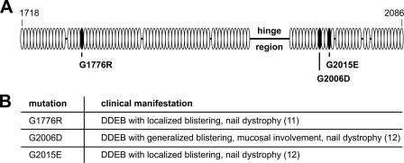
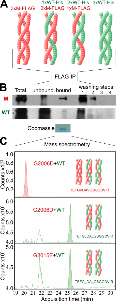
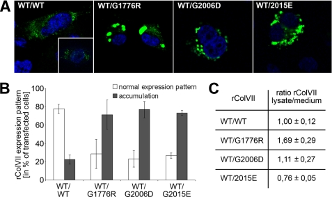
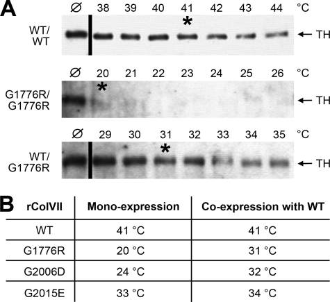
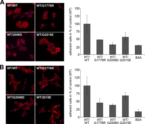
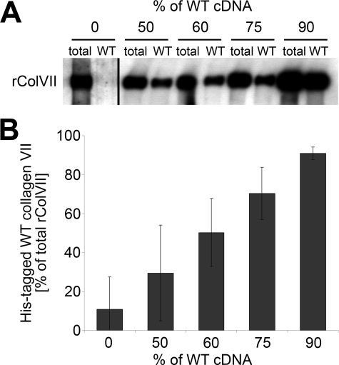
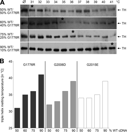
Dominant-negative interference by glycine substitution mutations in the COL7A1 gene causes dominant dystrophic epidermolysis bullosa (DDEB), a skin fragility disorder with mechanically induced blistering. Although qualitative and quantitative alterations of the COL7A1 gene product, collagen VII, underlie DDEB, the lack of direct correlation between mutations and the clinical phenotype has rendered DDEB less amenable to therapeutic targeting. To delineate the molecular mechanisms of DDEB, we used recombinant expression of wild-type (WT) and mutant collagen VII, which contained a naturally occurring COL7A1 mutation, G1776R, G2006D, or G2015E, for characterization of the triple helical molecules. The mutants were co-expressed with WT in equal amounts and could form heterotrimeric hybrid triple helices, as demonstrated by affinity purification and mass spectrometry. The thermal stability of the mutant molecules was strongly decreased, as evident in their sensitivity to trypsin digestion. The helix-to-coil transition, T(m), of the mutant molecules was 31-34 degrees C, and of WT collagen VII 41 degrees C. Co-expression of WT with G1776R- or G2006D-collagen VII resulted in partial intracellular retention of the collagen, and mutant collagen VII had reduced ability to support cell adhesion. Intriguingly, controlled overexpression of WT collagen VII gradually improved the thermal stability of the collective of collagen VII molecules. Co-expression in a ratio of 90% WT:10% mutant increased the T(m) to 41 degrees C for G1776R-collagen VII and to 39 degrees C for G2006D- and G2015E-collagen VII. Therefore, increasing the expression of WT collagen VII in the skin of patients with DDEB can be considered a valid therapeutic approach.


Figures

FIGURE 1.
FIGURE 1.
Position of glycine substitution mutations G1776R, G2006D, and G2015E and their clinical phenotype. A, schematic representation of the mutations included in this study and their location within the triple helical domain of collagen VII. Shown is the stretch of amino acids 1718–2086 around the hinge region in the central triple helix. Open ovals, Gly-X-Y repeats; black ovals, Gly-X-Y with a glycine substitution mutation; black bar, interruptions of the Gly-X-Y repeat pattern. B, phenotype in patients carrying one COL7A1 allele with the respective mutation.
FIGURE 2.
FIGURE 2.
Demonstration of WT:mutant hybrid triple helices using affinity purification and mass spectrometry. To demonstrate the formation of WT:mutant hybrid triple helices, His-tagged WT collagen VII and FLAG-tagged G1776R-L102/105V-, G2006D-L102/105V-, and G2015E-L102/105V collagen VII were co-expressed in HEK293T cells using equal amounts of the respective cDNAs. After 48 h, conditioned medium was collected and FLAG tag containing collagen VII triple helices were isolated by passing the medium over an anti-FLAG M2-agarose column and, after extensive washing, eluting the bound fraction with FLAG peptide. Presence of His-tagged WT-collagen VII and FLAG-tagged mutant collagen VII in the input, flow-through, eluate, and washing buffer was analyzed by immunoblotting using a FLAG or His tag-specific antibody. The eluate of the M2-agarose was also subjected to mass spectrometry with multiple reaction monitoring. A, schematic representation of the theoretically possible triple helices after co-expression of WT and mutant collagen VII. Red, FLAG-tagged mutant collagen VII (M-FLAG); green, His-tagged WT collagen VII (WT-His). B, immunoblot of FLAG-tagged G2006D-L102/105V collagen VII (upper panel) and His-tagged WT (lower panel) collagen VII after separation by M2-agarose column. Both FLAG-tagged mutant and His-tagged WT collagen VII are detectable in the fraction bound to the anti-FLAG M2-agarose, indicating the presence of hybrid triple helices. M, FLAG-tagged G2006D-L102/105V-collagen VII detected by anti-FLAG antibodies; WT, His-tagged WT-collagen VII detected by anti-His antibodies; Total, medium before incubation with M2-agarose; Unbound, medium after incubation with M2-agarose; Bound, fraction after elution of the column with FLAG peptide; washing steps 1–4, collagen VII in the subsequent washing steps before elution. C, collagen VII eluted from the M2-agarose was isolated by SDS-PAGE, and the Coomassie Blue-stained band was subjected to in-gel tryptic digest. Mass spectrometry with multiple reaction monitoring detected the presence of the peptide sequences TEFGVDAVGSGGDVIR and TEFGLDALGSGGDVIR representing mutant and WT collagen VII, respectively.
FIGURE 3.
FIGURE 3.
Intracellular accumulation of collagen VII containing mutations G1776R, G2006D, or G2015E. A, transfection of immortalized collagen VII-deficient keratinocytes with WT collagen VII alone shows an expression pattern (left panel) comparable to primary human keratinocytes (inset). Untransfected collagen VII-deficient keratinocytes remained completely negative (not shown). Co-transfection with cDNA for WT and mutant collagen VII in equal ratios leads to intracellular accumulation of collagen VII in case of glycine substitution G1776R (second panel from the left), glycine substitution G2006D (second panel from the right), and glycine substitution G2015E (right panel). Collagen VII is visualized by immunostaining with the NC2–10 antibody (green), nuclei are counterstained with DAPI (blue). B, percentage of transfected cells showing a normal expression pattern (white column) or intracellular accumulation (black column). C, quantification of rColVII in lysates and medium of transfected cells. Values are given as a ratio of rColVII in the cell lysate to rColVII in the medium. For comparison, the ratio observed after transfection with WT collagen VII was defined as 1.0. The data represent averages of at least two independent experiments.
FIGURE 4.
FIGURE 4.
Thermal stability of WT and mutant collagen VII. The thermal stability of the collagen triple helix of WT and mutant collagen VII was determined by limited proteolytic digestion. Collagen VII precipitated from the medium of transiently transfected HEK-293T cells was first subjected to pepsin digestion to remove the non-collagenous domains. After exposure to temperatures from 20 to 44 °C, the triple helices were subjected to limited trypsin digestion. The amount of rColVII resistant to tryptic digestion was determined by immunoblotting with the NC2–10 antibody. The temperature, at which 50% of the rColVII molecules were sensitive to proteolytic degradation was defined as “melting temperature, Tm”. A, sensitivity of WT rColVII to tryptic digestion (upper panel). The Tm of 41 °C (labeled with asterisk) is in agreement with previous data obtained by digesting native collagen VII produced by normal human keratinocytes (15). Mono-expression of G1776R-collagen VII resulted in a Tm below 20 °C (middle panel, Tm labeled with asterisk), whereas hybrid rColVII obtained by co-transfection of WT and G1776R-collagen VII cDNA shows a Tm of 31 °C (lower panel, Tm labeled with asterisk). ℘, collagen VII digested with trypsin at 20 °C; TH, migration position of the triple helix. B, overview of the melting temperatures of WT and mutant rColVII with the mutations G1776R, G2006D, and G2015E in mono- and co-expression with WT collagen VII, respectively.
FIGURE 5.
FIGURE 5.
Adhesion and spreading of normal human keratinocytes and fibroblasts on WT and mutant collagen VII. For these experiments, FLAG-tagged WT and hybrid collagen VII composed of WT and mutant rColVII was produced by co-transfecting HEK293 cells with the corresponding cDNA. After FLAG tag affinity purification, the collagens were coated onto coverslips in a concentration of 20 μg/ml; bovine serum albumin coating was used as negative control. Normal human keratinocytes or fibroblasts were incubated on the coverslips for 2 h, washed, fixed, and stained with DAPI and rhodamin-coupled phalloidin. A, spreading (left panel) and adhesion (right panel) of normal human keratinocytes on WT collagen VII and on the mutants G1776R, G2006D, or G2015E. B, spreading (left panel) and adhesion (right panel) of normal human fibroblasts on WT collagen VII and the mutants G1776R, G2006D, or G2015E. All three collagen VII mutants caused decreased adhesion and abnormal spreading of both cell types. The number of attached cells was evaluated by counting the DAPI-stained nuclei (in blue). The spreading was visualized by staining the actin cytoskeleton (in red). Shown are representative data of three independent experiments.
FIGURE 6.
FIGURE 6.
Quantification of mutant and WT collagen VII after co-transfection. HEK-293T cells were transiently transfected with a mixture of plasmid cDNA for His-tagged WT and FLAG-tagged mutant collagen VII. The amount of WT collagen VII in the culture medium was determined by quantitatively binding the FLAG-tagged mutant collagen VII to anti-FLAG-agarose, followed by quantification of the remaining His-tagged WT collagen VII in the unbound fraction using the NC2–10 antibody. A, immunoblot of collagen VII after incubation of hybrid molecules of His-tagged WT rColVII and FLAG-tagged mutant rColVII with an anti-FLAG column. Increasing the ratio of WT/mutant collagen VII cDNA in the transfection mix correlated with the amounts of collagen VII in the medium unable to bind to the anti-FLAG-agarose. Total, unbound fraction after incubation of medium with empty agarose; WT, unbound fraction after incubation of the medium with anti-FLAG-agarose. B, densitometric quantification of three independent immunoblots assaying the amount of His-tagged WT collagen VII after transient co-transfection of HEK-293T cells with mixtures of His-tagged WT and FLAG-tagged mutant collagen VII. Shown is the amount of His-tagged WT collagen VII in relation to the total rColVII.
FIGURE 7.
FIGURE 7.
Controlled overexpression of WT collagen VII improves triple helix stability of collagen VII containing glycine substitutions. Mutant collagen VII with glycine substitutions G1776R, G2006D, or G2015E was co-expressed with increasing amounts of WT collagen VII. For determination of the thermal stability, the hybrid molecules in the medium were subjected to limited proteolytic digestion and immunoblotting with collagen VII antibodies. A, when WT collagen VII and the mutant G1776R were co-expressed in equal amounts, 50% of the hybrid molecules were sensitive to proteolysis at 31 °C (marked by asterisk). Co-expression of 60% WT collagen VII and 40% of the mutant increased the Tm to 35 °C (asterisk). Co-expression of 75% WT collagen VII and 25% mutant lead to a Tm of 36 °C (asterisk), and co-expression of 90% WT collagen VII and 10% mutant to a Tm of 41 °C (asterisk). ℘, collagen VII digested with trypsin at 20 °C; TH, triple helix. Asterisks mark the melting temperature. B, correlation of thermal stability of hybrid collagen VII composed of increasing amounts of WT and decreasing amounts of mutant collagen VII. The thermal stability of all three mutants was clearly augmented by co-expression with increasing amounts of WT cDNA.

References

    1. Kern J. S., Has C. (2008) Expert. Rev. Dermatol. 3, 721–733
    1. Fine J. D., Eady R. A., Bauer E. A., Bauer J. W., Bruckner-Tuderman L., Heagerty A., Hintner H., Hovnanian A., Jonkman M. F., Leigh I., McGrath J. A., Mellerio J. E., Murrell D. F., Shimizu H., Uitto J., Vahlquist A., Woodley D., Zambruno G. (2008) J. Am. Acad. Dermatol. 58, 931–950 - PubMed
    1. Rattenholl A., Pappano W. N., Koch M., Keene D. R., Kadler K. E., Sasaki T., Timpl R., Burgeson R. E., Greenspan D. S., Bruckner-Tuderman L. (2002) J. Biol. Chem. 277, 26372–26378 - PubMed
    1. Burgeson R. E. (1993) J. Invest. Dermatol. 101, 252–255 - PubMed
    1. Kadler K. E., Baldock C., Bella J., Boot-Handford R. P. (2007) J. Cell Sci. 120, 1955–1958 - PubMed

Publication types