. 2009 Oct 30;284(44):30240-7.
doi: 10.1074/jbc.M109.046391. Epub 2009 Sep 2.

Highly conserved cysteines within the Ly6 domain of GPIHBP1 are crucial for the binding of lipoprotein lipase

Affiliations

Highly conserved cysteines within the Ly6 domain of GPIHBP1 are crucial for the binding of lipoprotein lipase

Anne P Beigneux et al. J Biol Chem. .

Abstract

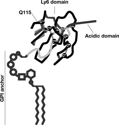
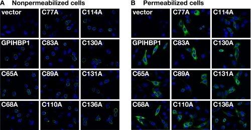
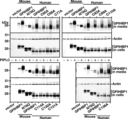
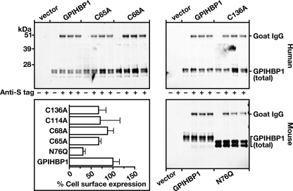
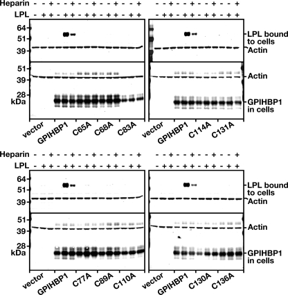
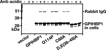
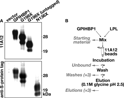
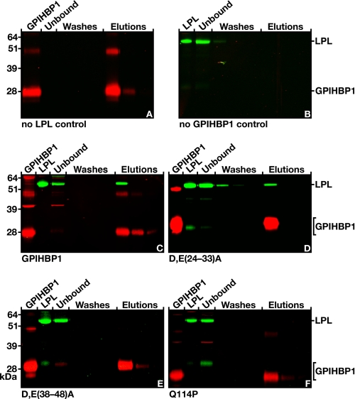
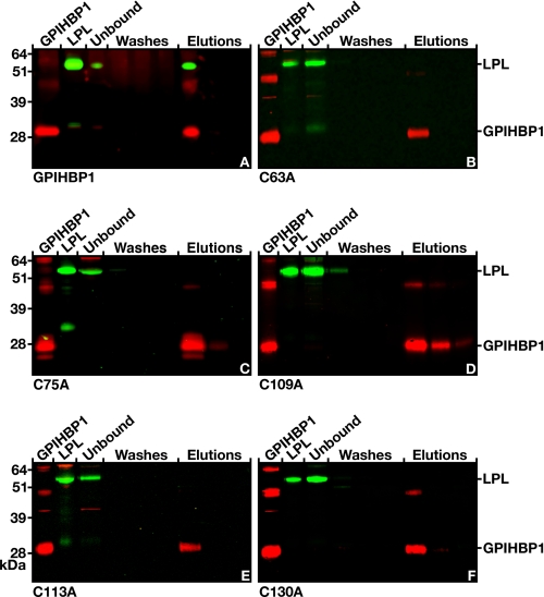
GPIHBP1, a glycosylphosphatidylinositol-anchored endothelial cell protein of the lymphocyte antigen 6 (Ly6) family, binds lipoprotein lipase (LPL) avidly and is required for the lipolytic processing of triglyceride-rich lipoproteins. GPIHBP1 contains two key structural motifs, an acidic domain and an Ly6 motif (a three-fingered domain specified by 10 cysteines). The acidic domain is required for LPL binding, but the importance of the Ly6 domain is less clear. To explore that issue, we transfected cells with a wild-type GPIHBP1 expression vector or mutant GPIHBP1 vectors in which specific cysteines in the Ly6 domain were changed to alanine. The mutant GPIHBP1 proteins reached the cell surface, as judged by antibody binding studies and by the ability of a phosphatidylinositol-specific phospholipase C to release these proteins from the cell surface. However, cells expressing the cysteine mutants could not bind LPL. The acidic domain of the cysteine mutants appeared to remain accessible, as judged by binding studies with an antibody against the acidic domain. We also developed a cell-free assay of LPL binding. We created a rat monoclonal antibody against the carboxyl terminus of mouse GPIHBP1 and used that antibody to coat agarose beads. We then tested the ability of soluble forms of GPIHBP1 that had been immobilized on monoclonal antibody-coated beads to bind LPL. In this assay, wild-type soluble GPIHBP1 bound LPL avidly, but the cysteine mutants did not. Thus, our studies suggest that a structurally intact Ly6 domain (in addition to the acidic domain) is essential for LPL binding.


Figures

FIGURE 1.
FIGURE 1.
Schematic of human GPIHBP1, showing the location of the acidic domain and the 10 highly conserved cysteines of the Ly6 domain. The location of Gln115 is also shown; a Q115P mutation was identified in association with chylomicronemia in a young man (15). This figure is modified, with permission, from a figure published in Ref. .
FIGURE 2.
FIGURE 2.
Mutant GPIHBP1 proteins with cysteine-to-alanine mutations within the Ly6 domain reach the cell surface. CHO pgsA-745 cells were electroporated with an empty vector, a construct encoding an S-protein-tagged human GPIHBP1, or mutant GPIHBP1 constructs in which specific cysteines within the Ly6 domain were replaced with alanine. The presence of GPIHBP1 in nonpermeabilized (A) and permeabilized (B) cells was assessed by immunofluorescence microscopy with a goat antiserum against the S-protein tag (green). The cell nuclei were visualized with 4′,6-diamidino-2-phenylindole (blue).
FIGURE 3.
FIGURE 3.
Mutant GPIHBP1 proteins with cysteine-to-alanine mutations within the Ly6 domain can be released from the cell surface with a PIPLC. CHO pgsA-745 cells were electroporated with expression vectors encoding wild-type mouse GPIHBP1, a mutant mouse GPIHBP1 (N76Q) that eliminates the sole N-linked glycosylation site, wild-type human GPIHBP1, and mutant human GPIHBP1 proteins with specific cysteine-to-alanine mutations in the Ly6 domain. All of the GPIHBP1 constructs contained an S-protein tag. Amounts of GPIHBP1 in the medium were assessed by Western blotting, both in untreated cells and in cells that had been treated with PIPLC (5 units/ml for 1 h at 37 °C).
FIGURE 4.
FIGURE 4.
Assessing relative amounts of GPIHBP1 on the surface of cells with an antiserum against the S-protein tag. CHO pgsA-745 cells were electroporated with vectors encoding wild-type mouse GPIHBP1, a mutant mouse GPIHBP1 (N76Q) in which the sole N-linked glycosylation site was eliminated, wild-type human GPIHBP1, and mutant human GPIHBP1 proteins with specific cysteine-to-alanine mutations in the Ly6 domain. All of the GPIHBP1 constructs contained an S-protein tag. 24 h after the electroporation, the medium was removed, and the cells were incubated for 2 h at 4 °C with a goat antibody against the S-protein tag. After removing the antibody, the cells were washed six times with ice-cold PBS. The cell extracts were prepared, and Western blots were performed with an anti-goat secondary antibody and either a rat monoclonal against mouse GPIHBP1 (in the case of cells transfected with mouse GPIHBP1 constructs) or a mouse monoclonal against human GPIHBP1 (in the case of cells transfected with human GPIHBP1 constructs). Band intensities were quantified with an Odyssey infrared scanner (Li-Cor). The signal corresponding to the goat anti-S-protein tag IgG was normalized to the binding of the rat antibody against mouse GPIHBP1 (for experiments with mouse GPIHBP1) or the monoclonal antibody against human GPIHBP1 (for experiments with human GPIHBP1) and expressed relative to wild-type control (set at 100%).
FIGURE 5.
FIGURE 5.
Reduced binding of human LPL to cells expressing mutant versions of GPIHBP1 with specific cysteine-to-alanine mutations in the Ly6 domain. CHO pgsA-745 cells were electroporated with an S-protein-tagged wild-type GPIHBP1 construct or mutant constructs with specific cysteine-to-alanine mutations. 24 h after the electroporation, the cells were incubated with V5-tagged human LPL in the presence or absence of heparin (500 units/ml). After washing the cells six times, the cell extracts were prepared, and the level of LPL bound to the cells was assessed by Western blotting with a V5 tag-specific antibody. Simultaneously, the level of GPIHBP1 in cell extracts was assessed by Western blotting with an antibody against the S-protein tag. Actin was used as a loading control.
FIGURE 6.
FIGURE 6.
Mutations in mouse GPIHBP1 that interfere with LPL binding (Q114P and C88A) do not appear to interfere with the accessibility of antibodies against the acidic domain of GPIHBP1. CHO pgsA-745 cells were electroporated with vectors encoding wild-type mouse GPIHBP1, GPIHBP1 with a Q114P mutation, GPIHBP1 with a C88A mutation, and GPIHBP1 in which all of the aspartates and glutamates between amino acids 38 and 48 were replaced with alanines (D/E(38–48)A). All of the GPIHBP1 constructs contained an S-protein tag. The next day the medium was removed, and the cells were incubated for 2 h at 4 °C with a rabbit polyclonal antibody raised against a peptide spanning the entire acidic domain of mouse GPIHBP1 (17). After removing the antibody, the cells were washed six times with ice-cold PBS. The cell extracts were prepared for Western blots with a goat antibody against the S-protein tag (to detect total GPIHBP1 in cells) and a donkey anti-rabbit secondary antibody (to detect binding of the anti-acidic domain rabbit antibody).
FIGURE 7.
FIGURE 7.
Schematic of a new method to assess the binding of LPL to soluble mouse GPIHBP1 captured on monoclonal antibody 11A12-coated agarose beads. A, Western blots to characterize antibody 11A12, a new rat monoclonal antibody against mouse GPIHBP1. The following GPIHBP1 proteins were examined: an S-protein-tagged GPIHBP1, S-protein-tagged and untagged “soluble” GPIHBP1 (G198X, truncated immediately before the GPI anchoring site), and S-protein-tagged “truncated” GPIHBP1 (N136X, truncated immediately after the Ly6 domain). Western blots were performed with antibody 11A12 and a goat antibody against an S-protein tag (located at the amino terminus of GPIHBP1 proteins); the binding of antibody 11A12 and the goat anti-S protein tag antibody was detected with donkey anti-rat (top) and anti-goat (bottom) secondary antibodies, respectively. B, a new monoclonal antibody-based assay to assess the binding of LPL to GPIHBP1. Agarose beads were coated with monoclonal antibody 11A12 as described under “Experimental Procedures.” The beads were then incubated with S-protein-tagged soluble GPIHBP1 and V5-tagged human LPL (from concentrated cell culture medium). The beads were washed with the incubation buffer, and the GPIHBP1 was eluted from the beads with 0.1 m glycine, pH 2.5. Western blots were performed to detect soluble GPIHBP1 and LPL in the following fractions: the samples loaded onto the beads (Starting material), the Unbound fraction, three sequential washes of the beads, and three sequential elutions of the beads with 0.1 m glycine, pH 2.5.
FIGURE 8.
FIGURE 8.
Testing the ability of human LPL to bind to soluble mouse GPIHBP1 that had been captured by antibody 11A12-coated agarose beads. A, binding of soluble GPIHBP1 alone (no LPL control) to antibody 11A12-coated beads. The soluble GPIHBP1 was eluted with 0.1 m glycine, pH 2.5. B, absence of LPL binding to antibody 11A12-coated beads (no GPIHBP1 control). C, binding of LPL to soluble wild-type GPIHBP1 that had been captured on antibody 11A12-coated beads; GPIHBP1 and LPL were both released with 0.1 m glycine, pH 2.5. D, binding of LPL to a mutant version of soluble GPIHBP1 (D/E(24–33)A) (17) that had been captured on antibody 11A12-coated beads. In D/E(24–33)A, all of the aspartates and glutamates between residues 24 and 33 were changed to alanine. E, absence of LPL binding to a mutant version of soluble GPIHBP1 (D/E(38–48)A) (17) that had been captured on antibody 11A12-coated beads. In D/E(38–48)A, all of the aspartates and glutamates between residues 38 and 48 were changed to alanine. F, absence of LPL binding to a mutant version of soluble GPIHBP1 (Q114P) (15) that had been captured on antibody 11A12-coated beads.
FIGURE 9.
FIGURE 9.
Testing the ability of human LPL to bind to cysteine mutant forms of GPIHBP1 captured by monoclonal antibody 11A12-coated agarose beads. A, Western blot showing the binding of LPL to wild-type soluble GPIHBP1 that had been immobilized on antibody 11A12-coated beads. GPIHBP1 and LPL were both released by 0.1 m glycine, pH 2.5. B–F, Western blots showing the absence of LPL binding to mutant soluble GPIHBP1 proteins harboring specific cysteine-to-alanine mutations within the Ly6 domain of GPIHBP1. Structural studies of three Ly6 proteins has revealed that the first cysteine forms a disulfide bond with the fifth cysteine, the second with the third, the fourth with the sixth, the seventh with the eight, and the ninth with the tenth (–10). The five cysteine mutants (C63A, C75A, C109A, C113A, and C130A) shown here were predicted to interrupt five different disulfide bonds in GPIHBP1 (10). GPIHBP1 mutants corresponding to the opposite cysteine in the disulfide pair (C66A, C81A, C88A, C129A, and C135A) were also created and tested in the monoclonal antibody-based assay; none of these mutants bound LPL (see supplemental Fig. 2).

References

    1. Beigneux A. P., Davies B. S., Gin P., Weinstein M. M., Farber E., Qiao X., Peale P., Bunting S., Walzem R. L., Wong J. S., Blaner W. S., Ding Z. M., Melford K., Wongsiriroj N., Shu X., de Sauvage F., Ryan R. O., Fong L. G., Bensadoun A., Young S. G. (2007) Cell Metab. 5, 279–291 - PMC - PubMed
    1. Ioka R. X., Kang M. J., Kamiyama S., Kim D. H., Magoori K., Kamataki A., Ito Y., Takei Y. A., Sasaki M., Suzuki T., Sasano H., Takahashi S., Sakai J., Fujino T., Yamamoto T. T. (2003) J. Biol. Chem. 278, 7344–7349 - PubMed
    1. Hata A., Ridinger D. N., Sutherland S., Emi M., Shuhua Z., Myers R. L., Ren K., Cheng T., Inoue I., Wilson D. E., Iverius P. H., Lalouel J. M. (1993) J. Biol. Chem. 268, 8447–8457 - PubMed
    1. Ma Y., Henderson H. E., Liu M. S., Zhang H., Forsythe I. J., Clarke-Lewis I., Hayden M. R., Brunzell J. D. (1994) J. Lipid Res. 35, 2049–2059 - PubMed
    1. Sendak R. A., Bensadoun A. (1998) J. Lipid Res. 39, 1310–1315 - PubMed

Publication types

LinkOut - more resources