

Single-molecule analysis of proteinxDNA complexes formed during partition of newly replicated plasmid molecules in Streptococcus pyogenes
- PMID: 19726689
- PMCID: PMC2781585
- DOI: 10.1074/jbc.M109.035410
Single-molecule analysis of proteinxDNA complexes formed during partition of newly replicated plasmid molecules in Streptococcus pyogenes
Abstract
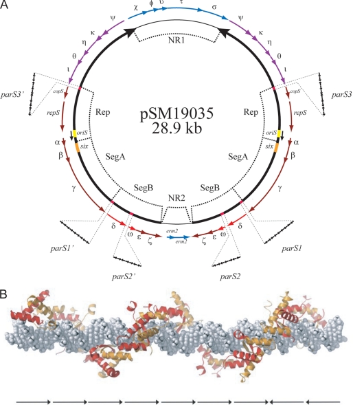
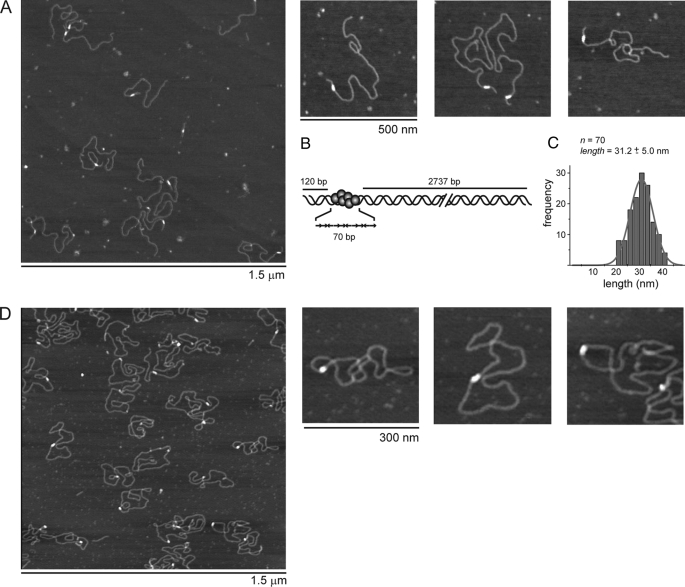
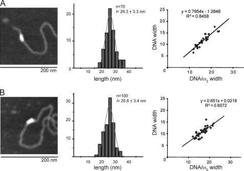
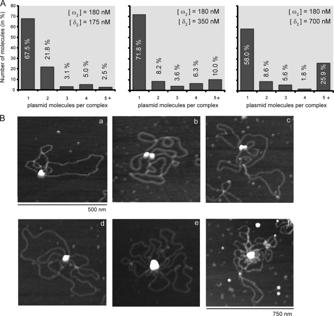
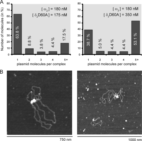
The Streptococcus pyogenes pSM19035 partition locus is ubiquitous among plasmids from vancomycin- or methicillin-resistant bacteria. An increasing understanding of this segregation system may highlight novel protein targets that could be blocked to curb bacterial proliferation. pSM19035 segregation depends on two homodimeric (delta(2) (ParA) and omega(2) (ParB)) proteins and six cis-acting centromeric noncurved parS sites. In the presence of ATPxMg(2+), delta(2) (delta x ATP x Mg(2+))(2) binds DNA in a sequence-independent manner. Protein omega(2) binds with high affinity and cooperatively to B-form parS DNA. Atomic force microscopy experiments indicate that about 10 omega(2) molecules bind parS, consisting of 10 contiguous iterons. Protein (delta x ATP x Mg(2+))(2), by interacting with the N terminus of omega(2) bound to parS, loses its association with DNA and relocalizes with omega(2).parS to form a ternary complex ((deltaxATPxMg(2+))(2) x omega(2) x parS) with the DNA remaining in straight B-form. Then, the interaction of two (delta x ATP x Mg(2+))(2).omega(2).parS complexes via delta(2) promotes pairing of a plasmid subfraction. (deltaD60A x ATP x Mg(2+))(2), which binds but does not hydrolyze ATP, leads to accumulation of pairing intermediates, suggesting that ATP hydrolysis induces plasmid separation. We propose that the molar omega(2):delta(2) ratio regulates the different stages of pSM19035 segregation, pairing, and delta(2) polymerization, before cell division.
Figures
Similar articles
-
Streptococcus pyogenes pSM19035 requires dynamic assembly of ATP-bound ParA and ParB on parS DNA during plasmid segregation.Nucleic Acids Res. 2008 Jun;36(11):3676-89. doi: 10.1093/nar/gkn170. Epub 2008 May 13. Nucleic Acids Res. 2008. PMID: 18477635 Free PMC article.
-
Mapping of the interactions between partition proteins Delta and Omega of plasmid pSM19035 from Streptococcus pyogenes.Microbiology (Reading). 2011 Apr;157(Pt 4):1009-1020. doi: 10.1099/mic.0.045369-0. Epub 2011 Jan 20. Microbiology (Reading). 2011. PMID: 21252276
-
ParAB Partition Dynamics in Firmicutes: Nucleoid Bound ParA Captures and Tethers ParB-Plasmid Complexes.PLoS One. 2015 Jul 10;10(7):e0131943. doi: 10.1371/journal.pone.0131943. eCollection 2015. PLoS One. 2015. PMID: 26161642 Free PMC article.
-
The Interplay between Different Stability Systems Contributes to Faithful Segregation: Streptococcus pyogenes pSM19035 as a Model.Microbiol Spectr. 2014 Aug;2(4):PLAS-0007-2013. doi: 10.1128/microbiolspec.PLAS-0007-2013. Microbiol Spectr. 2014. PMID: 26104212 Review.
-
Plasmid pSM19035, a model to study stable maintenance in Firmicutes.Plasmid. 2010 Jul;64(1):1-17. doi: 10.1016/j.plasmid.2010.04.002. Epub 2010 Apr 18. Plasmid. 2010. PMID: 20403380 Review.
Cited by
-
Distinct architectural requirements for the parS centromeric sequence of the pSM19035 plasmid partition machinery.Elife. 2022 Sep 5;11:e79480. doi: 10.7554/eLife.79480. Elife. 2022. PMID: 36062913 Free PMC article.
-
RecA Regulation by RecU and DprA During Bacillus subtilis Natural Plasmid Transformation.Front Microbiol. 2018 Jul 11;9:1514. doi: 10.3389/fmicb.2018.01514. eCollection 2018. Front Microbiol. 2018. PMID: 30050509 Free PMC article.
-
Single molecule analysis reveals monomeric XPA bends DNA and undergoes episodic linear diffusion during damage search.Nat Commun. 2020 Mar 13;11(1):1356. doi: 10.1038/s41467-020-15168-1. Nat Commun. 2020. PMID: 32170071 Free PMC article.
-
Role of vimA in cell surface biogenesis in Porphyromonas gingivalis.Microbiology (Reading). 2010 Jul;156(Pt 7):2180-2193. doi: 10.1099/mic.0.038331-0. Epub 2010 Apr 8. Microbiology (Reading). 2010. PMID: 20378652 Free PMC article.
-
VimA-dependent modulation of the secretome in Porphyromonas gingivalis.Mol Oral Microbiol. 2012 Dec;27(6):420-35. doi: 10.1111/j.2041-1014.2012.00661.x. Epub 2012 Aug 9. Mol Oral Microbiol. 2012. PMID: 23134608 Free PMC article.
References
Publication types
MeSH terms
Substances
LinkOut - more resources
Full Text Sources
Miscellaneous
