. 2009 Sep;119(9):2737-44.
doi: 10.1172/JCI38292. Epub 2009 Aug 24.

Impaired endocytosis of the ion channel TRPM4 is associated with human progressive familial heart block type I

Affiliations

Impaired endocytosis of the ion channel TRPM4 is associated with human progressive familial heart block type I

Martin Kruse et al. J Clin Invest. 2009 Sep.

Abstract

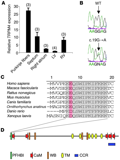
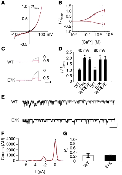
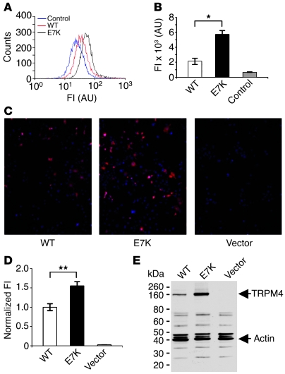
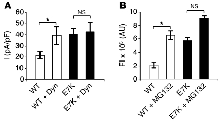
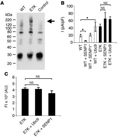
Progressive familial heart block type I (PFHBI) is a progressive cardiac bundle branch disease in the His-Purkinje system that exhibits autosomal-dominant inheritance. In 3 branches of a large South African Afrikaner pedigree with an autosomal-dominant form of PFHBI, we identified the mutation c.19G-->A in the transient receptor potential cation channel, subfamily M, member 4 gene (TRPM4) at chromosomal locus 19q13.3. This mutation predicted the amino acid substitution p.E7K in the TRPM4 amino terminus. TRPM4 encodes a Ca2+-activated nonselective cation (CAN) channel that belongs to the transient receptor potential melastatin ion channel family. Quantitative analysis of TRPM4 mRNA content in human cardiac tissue showed the highest expression level in Purkinje fibers. Cellular expression studies showed that the c.19G-->A missense mutation attenuated deSUMOylation of the TRPM4 channel. The resulting constitutive SUMOylation of the mutant TRPM4 channel impaired endocytosis and led to elevated TRPM4 channel density at the cell surface. Our data therefore revealed a gain-of-function mechanism underlying this type of familial heart block.


Figures

Figure 1
Figure 1. Cardiac phenotype of PFHBI patients.
Shown are electrocardiographic examples of PFHBI. (A) Sinus rhythm with a RBBB in an 8-year-old asymptomatic boy on a standard 12-lead ECG, with leads Std I, V1, and V6 shown. (B) Shown is 2:1 atrioventricular node block (atrial rate, 76 bpm; ventricular rate, 38 bpm) with a broad QRS complex on Holter monitoring in a 54-year-old man who had recently become symptomatic. ECGs were recorded at a 25 mm/s paper speed and 10 mm/mV signal amplitude.
Figure 2
Figure 2. TRPM4 missense mutation in exon 1 associated with PFHBI.
(A) Relative expression of TRPM4 transcripts in different tissues of nondiseased human heart was assayed by quantitative RT-PCR. TRPM4 mRNA expression levels were normalized to the level in left ventricle. Numbers of individual probes are shown in parentheses. Each experiment was done in triplicate. (B) Electropherograms show TRPM4 WT sequence and the heterozygous sequence change c.19G→A in the DNA of PFHBI-affected individuals. (C) Partial amino acid sequence alignment of TRPM4 N terminus among different species. Gray shading shows the conserved sequence motif; red shading highlights the glutamic acid substituted by lysine in TRPM4 associated with PFHBI. Numbering refers to the human sequence. (D) Diagram of TRPM4 topology and functional domains, with 6 membrane-spanning domains (TM) flanked by N- and C-terminal cytoplasmic sequences. PFHBI, PFHBI domain; CaM, Calmodulin-binding domain; WB, Walker B ATP-binding motif; CCR, coiled-coiled region. Figure part adapted with permission from Pflügers Archiv (19).
Figure 3
Figure 3. Expression of human TRPM4 and TRPM4E7K in HEK 293 cells.
Unless otherwise indicated, black traces denote TRPM4; red traces denote TRPM4E7K. (A) Normalized current-voltage relationship for TRPM4 and TRPM4E7K obtained from 250-ms voltage ramps measured in the whole-cell patch-clamp configuration from –120 to +100 mV. Holding potential was –60 mV. (B) Ca2+ dependence of TRPM4 current densities (I/Imax) obtained from voltage ramps measured at –80 and +80 mV (n = 7–16). [Ca2+]i, intracellular Ca2+. (C) AMP-PNP block of WT TRPM4 and TRPM4E7K current. Holding potential was 0 mV. Currents, elicited by a 250-ms pulse to +100 mV after a 500-ms pulse to –100 mV, were recorded before (0; black) and after application of 500 μM AMP-PNP (0.5; red) Scale bars: 200 ms, 0.5 nA. (D) Normalized current densities (I/Inorm) of TRPM4E7K expressed alone (n = 13) or in a 1:1 ratio with TRPM4 (WT/E7K, n = 6) obtained from voltage ramps measured at +40 and +80 mV. n = 16 (WT TRPM4). *P < 0.05 versus WT. (E) Single-channel currents recorded from inside-out patches at –100 mV. Scale bars: 1 s, 5 pA. (F) Histogram plots of TRPM4 and TRPM4E7K traces shown in E. (G) Po of TRPM4 and TRPM4E7K channel at +100 mV (n = 8–9).
Figure 4
Figure 4. Analysis of TRPM4 and TRPM4E7K protein density.
(A) FACS fluorogram of nonpermeabilized cells expressing untagged TRPM4 control, Myc-tagged TRPM4, or Myc-tagged TRPM4E7K labeled with FITC-conjugated Myc-specific antibody. FI, fluorescence intensity. (B) FACS analysis of fluorograms in A. n = 9 per group. (CE) Cells expressing empty vector control (Vector), FLAG-tagged TRPM4, or FLAG-tagged TRPM4E7K were fixed and permeabilized 48 hours after transfection. (C) Cells were stained with mouse FLAG-specific primary antibody and anti-mouse goat Alexa Fluor 546–coupled secondary antibody. Nuclei were stained with DAPI. Original magnification, ×20. (D) Immunofluorescence intensity of FLAG-tagged TRPM4E7K normalized to that of FLAG-tagged TRPM4–expressing cells (n = 60 per group). (E) SDS-PAGE of C-terminally FLAG-tagged TRPM4 and TRPM4E7K protein in cell lysates followed by Western blotting. Membranes were stained with mouse FLAG-specific antibody and simultaneously with rabbit actin-specific antibody for loading control. *P < 0.05, **P < 0.01.
Figure 5
Figure 5. Posttranslational regulation of TRPM4 and TRPM4E7K channel density.
(A) TRPM4 and TRPM4E7K channel, expressed together with or without dynamitin (Dyn) for 18 hours in HEK 293 cells. Current densities were obtained at +80 mV as described in Figure 3A. n = 7 (E7K + Dyn); 9 (WT and WT + Dyn); 10 (E7K). (B) HEK 293 cells expressing Myc-tagged TRPM4 or TRPM4E7K channel were incubated with or without 10 μM MG132. FACS analysis was performed as in Figure 4A. n = 3–9. *P < 0.05.
Figure 6
Figure 6. Sensitivity of TRPM4 current density to SUMOylation.
(A) C-terminally FLAG-tagged TRPM4 or TRPM4E7K protein pulled down on beads coated with FLAG-specific antibodies were subjected to SDS-PAGE and Western blotting. Blots were stained with rabbit SUMO-1–specific primary antibody and anti-mouse rabbit horseradish peroxidase–coupled secondary antibody. Arrow indicates SUMOylated TRPM4. (B) Same experiment as in Figure 5A, but TRPM4 channel was coexpressed with Ubc9, SENP1, or the inactive mutant SENP1-C603S (SENP1*). n = 6–13. (C) Myc-tagged TRPM4E7K was expressed in HEK 293 cells alone or together with Ubc9 or SENP1. At 24 hours after transfection, the surface density of Myc-tagged TRPM4E7K channel was assessed by FACS analysis, as described in Methods. n = 3 per group. *P < 0.05.

Comment in

References

    1. Josephson, M.E., Zimetbaum, P., Marchlinski, F.E., and Buxton, A.E. 1998. The bradyarrhythmias: disorders of sinus node function and AV conduction disturbances. InHarrison’s principles of internal medicine . A.S. Fauci, et al., editors. McGraw-Hill. New York, New York, USA. 1253–1261.
    1. Ector H., et al. The registry of the European Working Group on Cardiac Pacing (EWGCP). A working group of the European Society of Cardiology. Europace. 2000;2:251–255. doi: 10.1053/eupc.2000.0109. - DOI - PubMed
    1. Epstein A.E., et al. ACC/AHA/HRS 2008 Guidelines for Device-Based Therapy of Cardiac Rhythm Abnormalities: a report of the American College of Cardiology/American Heart Association Task Force on Practice Guidelines (Writing Committee to Revise the ACC/AHA/NASPE 2002 Guideline Update for Implantation of Cardiac Pacemakers and Antiarrhythmia Devices): developed in collaboration with the American Association for Thoracic Surgery and Society of Thoracic Surgeons. Circulation. 2008;117:e350–e408. doi: 10.1161/CIRCUALTIONAHA.108.189742. - DOI - PubMed
    1. Benson D.W. Genetics of atrioventricular conduction disease in humans. Anat. Rec. A. Discov. Mol. Cell. Evol. Biol. 2004;280:934–939. - PubMed
    1. Brink P.A., et al. Gene for progressive familial heart block type I maps to chromosome 19q13. Circulation. 1995;91:1633–1640. - PubMed

Publication types

MeSH terms