Review
. 2009 Dec;23(12):2233-41.
doi: 10.1038/leu.2009.175. Epub 2009 Sep 3.

Contribution of bone microenvironment to leukemogenesis and leukemia progression

Affiliations
Review

Contribution of bone microenvironment to leukemogenesis and leukemia progression

F Ayala et al. Leukemia. 2009 Dec.

Abstract

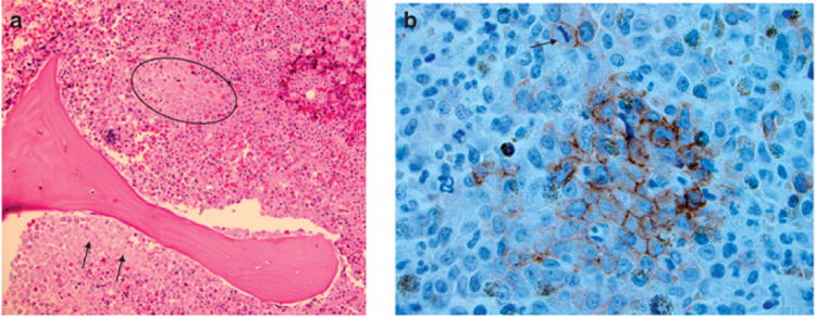
Tumor microenvironment has a major role in cancer progression and resistance to treatment. The bone marrow (BM) is a dynamic network of growth factors, cytokines and stromal cells, providing a permissive environment for leukemogenesis and progression. Both BM stroma and leukemic blasts promote angiogenesis, which is increased in acute lymphoblastic leukemia and acute myeloid leukemia. Growth factors like vascular endothelial growth factor (VEGF), basic fibroblast growth factor and angiopoietins are the main proangiogenic mediators in acute leukemia. Autocrine proleukemic loops have been described for VEGF and angiopoietin in hematopoietic cells. Interactions of stromal cells and extracellular matrix with leukemic blasts can also generate antiapoptotic signals that contribute to neoplastic progression and persistence of treatment-resistant minimal residual disease. High expression of CXC chemokine ligand 4 (CXCR4) by leukemic blasts and activation of the CXCR4-CXCL12 axis is involved in leukemia progression and disruption of normal hematopoiesis. Leukemia-associated bone microenvironment markers could be used as prognostic or predictive indicators of disease progression and/or treatment outcome. Studies related to bone microenvironment would likely provide a better understanding of the treatment resistance associated with leukemia therapy and design of new treatments.


Conflict of interest statement

Conflict of interest: The authors declare no conflict of interest.

Figures

Figure 1
Figure 1. Endovascular niche
(a) The endovascular niche is an area immediately surrounding vessels and vascular sinusoids. The endovascular niche is involved in regulation of hematopoietic elements, especially granulocytic and megakaryocytes. In many myeloid disorders (leukemia, myelodysplastic syndromes or myeloproliferative neoplasms) the vascular microvessel density is increased within the marrow stroma. A case of primary myelofibrosis is illustrated above, where there are abnormal clusters of megakaryocytes and increased sinusoids that are dilated. As shown in the lower (left) part of the photomicrograph, abnormal megakaryocytes are sometimes found within the vascular sinusoid (intra-sinusoidal hematopoiesis). This is not seen in a normal endovascular niche, where megakaryocytes are inhibited from crossing over the endothelial barrier. Normally, megakaryocytic cytoplasmic processes penetrate the endothelium and release platelets as a shower (thrombopoiesis). (b) In this case of advanced myeloproliferative neoplasm, because of an abnormal endovascular niche, immature elements gain access to peripheral blood through vascular sinusoids, as illustrated above. This includes myeloblasts resulting in a leukemic phase. Illustrated above is a cluster of immature erythroid cells within vascular sinusoids. Leukemia and tumor microenvironment
Figure 2
Figure 2. Endosteal niche and abnormal localization of immature precursors (ALIPS)
(a) Often seen in myelodysplastic syndrome ALIPS refers to a cluster of immature precursors away from its normal microenvironment (circle). Illustrated here is the localization of a cluster of myeloid precursors away from the normal endosteal niche. (b) CD117 stains highlight this cluster of immature precursors. A mitotic figure is also seen above the cluster (arrow).
Figure 3
Figure 3. Interactions between leukemic blasts and bone marrow stromal cells
Leukemic blasts and stromal cells produce vascular endothelial growth factor (VEGF), basic fibroblast growth factor (bFGF) and other proangiogenic mediators, which also have protumoral paracrine and autocrine effects. Integrins and CXCR4 are fundamental for blast–stroma adhesion and mediate blasts ‘homing’ and persistence of residual disease after treatment. Different mechanisms have been described for acute myeloid leukemia (AML) and acute lymphoblastic leukemia (ALL), the latter being more dependent on bFGF than on VEGF as proangiogenic mediator. ALL microenvironment is also richer in interleukin mediators and in asparaginase synthetase, another mechanism of resistance to treatment.

References

    1. Quesenberry PJ, Aliotta JM. The paradoxical dynamism of marrow stem cells: considerations of stem cells, niches and microvesicles. Stem Cell Rev. 2008;4:137–147. - PMC - PubMed
    1. Raaijmakers MHGP, Scadden DT. Evolving concepts on the micro environmental niche for hematopoietic stem cells. Curr Opin Hematol. 2008;15:301–306. - PubMed
    1. Lataillade JJ, Pierre-Louis O, Hasselbalch HC, Uzan G, Jasmin C, Martyré MC, et al. Does primary myelofibrosis involve a defective stem cell niche? From concept to evidence Blood. 2008;112:3026–3035. - PubMed
    1. Folkmann J, Kalluri R. Cancer without disease. Nature. 2008;427:787. - PubMed
    1. Nyberg P, Salo T, Kalluri R. Tumor microenvironment and angiogenesis. Front Biosci. 2008;13:6537–6553. - PubMed

Publication types

MeSH terms