

Case Reports
doi: 10.1007/s11999-009-0997-1.
Epub 2009 Sep 2.
Case report: Fresh-stored osteochondral allograft for treatment of osteochondritis dissecans the femoral head
Affiliations
- PMID: 19727986
- PMCID: PMC2806996
- DOI: 10.1007/s11999-009-0997-1
Item in Clipboard
Case Reports
Case report: Fresh-stored osteochondral allograft for treatment of osteochondritis dissecans the femoral head
Clin Orthop Relat Res.
2010 Feb.
Abstract
Osteochondral defects of the femoral head are exceedingly rare, with limited treatment options. Restoration procedures for similar defects involving the knee and ankle have been well described. In this report, we present a young patient who had a symptomatic osteochondral defect of the femoral head develop secondary to trauma and underwent subsequent treatment using a fresh-stored osteochondral allograft via a trochanteric osteotomy. At the 1-year followup, the patient was symptom free with near-complete incorporation of the graft radiographically. Our observations in this case suggest osteoarticular implantation may be an appropriate alternative to consider when treating osteochondral defects of the femoral head.
Figures

(A) Anteroposterior and (B) lateral radiographs reveal an osteochondral defect of the femoral head.

(A) Coronal and (B) sagittal T2-weighted MRI scans of the femoral head defect show the areas of high signal intensity are not contiguous and do not undermine the lesion, suggesting the fragment is stable without evidence of collapse.
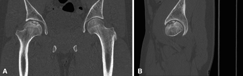
(A) Coronal and (B) sagittal CT scans show fragmentation of the lesion.

An intraoperative photograph shows the lesion involving the anterior superomedial aspect of the femoral head.

An intraoperative photograph shows the osteochondral plug after harvesting.
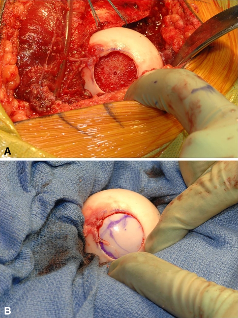
Intraoperative photographs illustrate (A) the host defect after preparation and (B) the osteochondral allograft after implantation.

Postoperative (A) anteroposterior and (B) lateral radiographs at 1-year followup show graft incorporation.
References
-
- Akimau P, Bhosale A, Harrison PE, Roberts S, McCall IW, Richardson JB, Ashton BA. Autologous chondrocyte implantation with bone grafting for osteochondral defect due to posttraumatic osteonecrosis of the hip: a case report. Acta Orthop. 2006;77:333–336. doi: 10.1080/17453670610046208. - DOI - PubMed
-
- Beaver RJ, Mahomed M, Backstein D, Davis A, Zukor DJ, Gross AE. Fresh osteochondral allografts for post-traumatic defects in the knee: a survivorship analysis. J Bone Joint Surg Br. 1992;74:105–110. - PubMed
Publication types
MeSH terms
LinkOut - more resources
Full Text Sources
Medical
