. 2009 Sep 3;16(1):81.
doi: 10.1186/1423-0127-16-81.

Myofibril-inducing RNA (MIR) is essential for tropomyosin expression and myofibrillogenesis in axolotl hearts

Affiliations

Myofibril-inducing RNA (MIR) is essential for tropomyosin expression and myofibrillogenesis in axolotl hearts

Chi Zhang et al. J Biomed Sci. .

Abstract

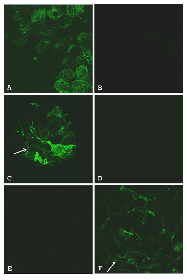
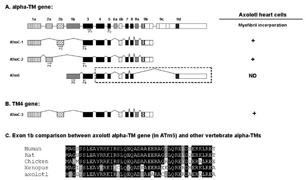
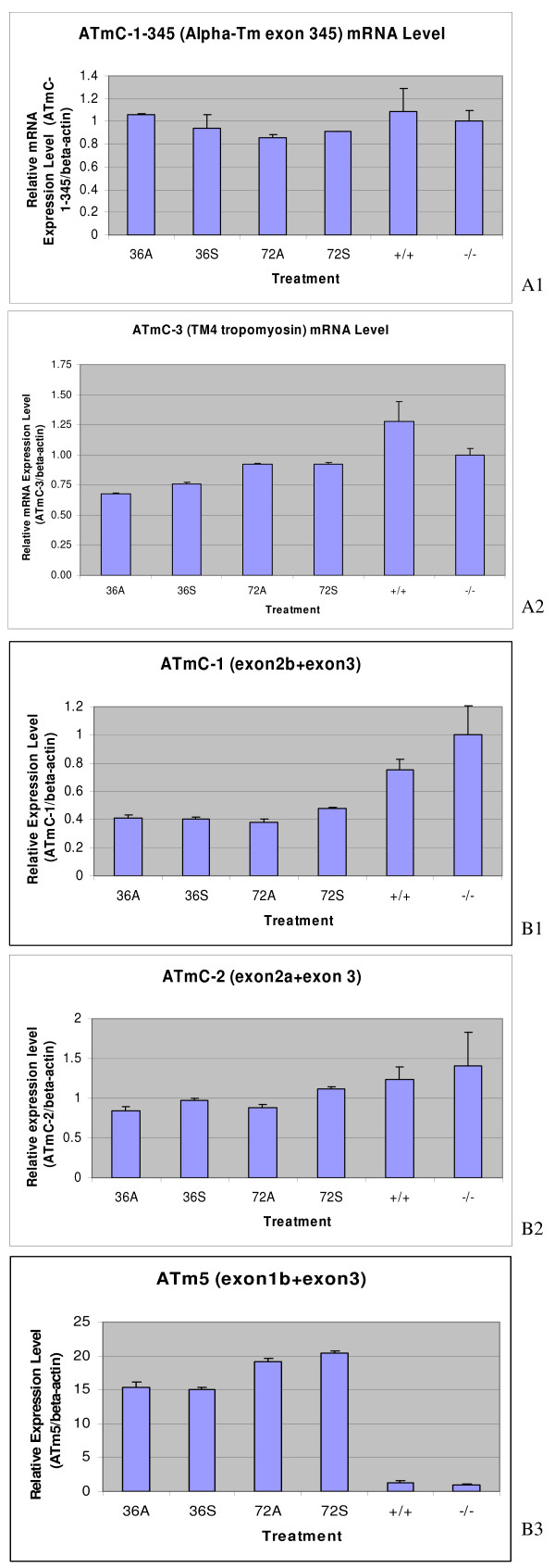
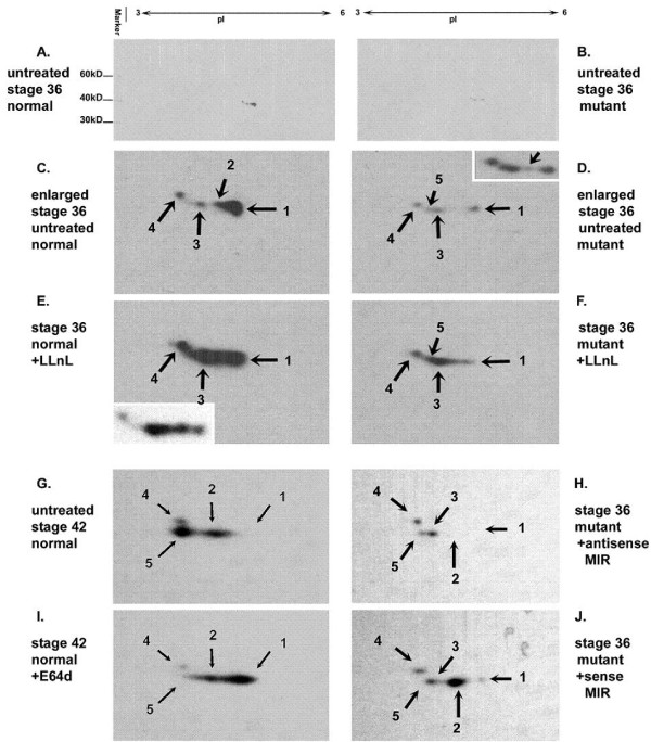
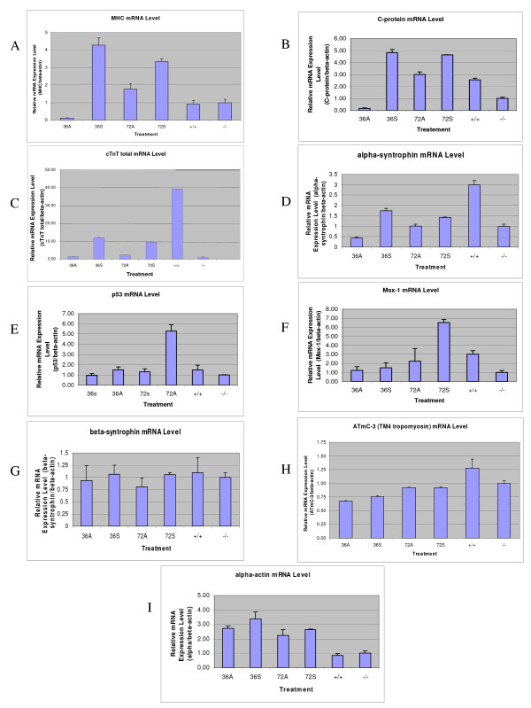
The Mexican axolotl, Ambystoma mexicanum, carries the naturally-occurring recessive mutant gene 'c' that results in a failure of homozygous (c/c) embryos to form hearts that beat because of an absence of organized myofibrils. Our previous studies have shown that a noncoding RNA, Myofibril-Inducing RNA (MIR), is capable of promoting myofibrillogenesis and heart beating in the mutant (c/c) axolotls. The present study demonstrates that the MIR gene is essential for tropomyosin (TM) expression in axolotl hearts during development. Gene expression studies show that mRNA expression of various tropomyosin isoforms in untreated mutant hearts and in normal hearts knocked down with double-stranded MIR (dsMIR) are similar to untreated normal. However, at the protein level, selected tropomyosin isoforms are significantly reduced in mutant and dsMIR treated normal hearts. These results suggest that MIR is involved in controlling the translation or post-translation of various TM isoforms and subsequently of regulating cardiac contractility.


Figures

Figure 1
Figure 1
The full length sequence of the MIR gene. Genomic DNA sequence: 1-379 bp (Underlined); Active MIR sequence (166 nt): 691-856 bp (Bold and underlined); Potential polyadenylation signals: 835-840 bp and 840-845 bp; Predicted TATA box: 343-350 bp (Shaded); Longest predicted open reading frame: 659-808 bp; Poly A+: 977-1000 bp.
Figure 2
Figure 2
Confocal microscopy of embryonic axolotl hearts stained with anti-tropomyosin antibody. Normal heart incubated with Steinberg's solution and lipofectin only has extensive tropomyosin staining (A). Mutant heart incubated with Steinberg's solution and lipofectin only has virtually no staining (B). Mutant heart incubated with Steinberg's solution, lipofectin and full length sense MIR shows significant tropomyosin (arrow) staining (C). Mutant heart incubated with Steinberg's solution, lipofectin and full length sense MIR pretreated with RNase. Very little tropomyosin is indicated in the cells (D). Mutant heart incubated with Steinberg's solution, lipofectin and full length antisense MIR. No staining for tropomyosin is observed (E). Mutant heart incubated with Steinberg's solution, lipofectin and 166 nt MIR RNA shows significant levels of tropomyosin (arrow) protein (F); Magnification: 60 ×.
Figure 3
Figure 3
Schematic representation of four different TM isoforms produced from alpha -TM (A) and TM4 type tropomyosin (B) genes. Each box represents an exon which is joined together by mRNA splicing. The alpha-TM gene is alternatively processed at both the 5' and/or 3' ends, as well as at the internal exons 6a/b. Our studies are focused on the 5' end sequence and exon 2a/b. The fibroblast type tropomyosin isoform (ATm5) has only the 5' sequence cloned with exon 1b and part of exon 3 cloned for RT-PCR studies, assuming the internal and 3' sequence are homologous to other vertebrates (dashed line box). Primers to amplify specific isoforms were designed based on the alternative spliced exons (P1 to P5). C. Highly conserved peptide sequence from exon 1b of axolotl alpha-tropomyosin compared to other vertebrate sequences. The indicated splicing pattern of axolotl tropomyosin genes are hypothetical based on other vertebrate sequences. ATmC-1, 2 and 3 were all demonstrated to be able to incorporate into myofibril structures [21].
Figure 4
Figure 4
Transcriptional levels of various tropomyosin isoforms (A1 and A2); Determination of mRNA splicing of various tropomyosin isoforms (B1, B2 and B3). Real-time RT-PCR showed no significant difference at the transcription level for alpha- (A1) and TM4 type tropomyosin (A2) genes in the mutant hearts treated with MIR sense or antisense RNA. 36A: Mutant hearts treated with antisense MIR for 36 hours; 36S: Mutant hearts treated with sense MIR for 36 hours; 72A: Mutant hearts treated with antisense MIR for 72 hours; 72S: Mutant hearts treated with sense MIR for 72 hours; +/+: homozygous normal hearts with no treatment; (-/-): homozygous mutant hearts with no treatment. Real-time RT-PCR showing no significant difference during mRNA splicing of the alpha-TM gene (the ATmC-1 containing exon 2b (B1), the ATmC-2 containing exon 2a (B2) and the fibroblast tropomyosin isoform with direct exon 1b and 3 conjunction (B3) in the mutant hearts treated with the MIR sense or MIR antisense.
Figure 5
Figure 5
Double-stranded MIR. Synthesized MIR double-stranded RNA loaded on 1.5% agarose gel for electrophoresis shows a 550 bp band. The size marker is loaded on the left-side of the gel.
Figure 6
Figure 6
Confocal microscopy of embryonic axolotl hearts treated with double-stranded MIR. The images show those hearts that stopped beating after 4 days but regained contractions later. Hearts were fixed after 9 days in culture. Normal heart (stage 36) organ cultured with lipofectin in Steinberg's Solution only for 10 days (A); Myofibril structures are clearly shown. Normal heart (stage 36) organ cultured with lipofectin in Steinberg's Solution with double-stranded RNA for 10 days (B); Mutant heart (stage 36) organ cultured with lipofectin in Steinberg's Solution with double-stranded RNA for 10 days (C). (Magnification: A-E: 60 ×; F and G: 10 ×). Hearts incubated with mock transfection (no dsRNA) show abundant staining of tropomyosin and organized myofibril structures (D) with nuclei stained by propidium iodide (PI). Hearts transfected by the dsRNA show a significant decrease of tropomyosin expression in more than half of the cells but continue to express in the remaining cells (E). Normal hearts treated with dsRNA for 9 days stained for tropomyosin (F). Clusters of cells with positive staining for tropomyosin are present in different areas. Normal hearts with mock transfection (no dsRNA) for 9 days stained for tropomyosin (G).
Figure 7
Figure 7
Myofibril morphological examinations. Normal embryonic hearts treated with double-stranded MIR for 4 days show a dramatic reduction in tropomyosin protein and myofibrils. Hearts scanned here with dsRNA transfection already have stopped beating at the time of fixation. Hearts incubated with mock transfection (no dsRNA) show abundant staining of tropomyosin and organized myofibril structures (A). Hearts transfected by dsRNA show almost no detectable expression of tropomyosin in the whole area of the ventricle (B). (Magnification: 60 ×).
Figure 8
Figure 8
Determination of myofibril protein expression. Real-time RT-PCR confirms the knockdown of endogenous MIR (A) in the normal hearts but shows no significant difference at the level of the transcripts of alpha- (B) and TM4 type tropomyosin (C) transcripts in the normal hearts treated with double-stranded MIR (dsRNA-) for 36 hours or 84 hours. However, both alpha- and TM4 type tropomyosin transcripts in the normal hearts treated with dsRNA for 9 days show dramatic decreases to nearly 1/3 of the levels in control groups with hearts incubated in lipofectamine transfection medium only. MIR expression in 9-day-cultured hearts is not detectable.
Figure 9
Figure 9
Two-dimensional SDS-PAGE of embryonic heart proteins followed by Western Blotting using CH1 (anti-tropomyosin) antibody. Untreated stage 36 normal hearts (A); Untreated stage 36 mutant hearts (B); Enlarged figure from A for CH1-recognizable tropomyosin isoforms in untreated normal hearts (C); Enlarged figure from B for CH1-recognizable tropomyosin isoforms in untreated normal hearts (D); The top right corner after overexposure of the same blot as D shows that mutant hearts are expressing isoform 2 as well as isoforms 1, 3 and 4. Mutant hearts at this stage are expressing all of the isoforms as normal hearts at much lower levels along with an extra isoform (5) which is detectable only at later developmental stages in normal hearts (G); Stage 36 normal hearts incubated with LLnL for 10 hours. The left bottom corner shows the less exposed image after Western blotting on the same sample, clearly showing increased protein concentration for isoform 2, 3 and 4 but not for 1 (E); Stage 36 mutant hearts incubated with LLnL for 10 hours. Increasing of protein concentration for isoform 3, 4 and 5 is clearly detected but not for 1 (F); Untreated stage 42 normal hearts (G); Stage 42 normal hearts incubated with E64d for 10 hours (I); Protection for isoform 1 is detected. Stage 36 mutant hearts treated by antisense MIR for 4 days (H); Stage 36 mutant hearts treated by sense MIR for 4 days (J); A significant increase in spot density for isoform 2 is detected when compared to untreated mutant hearts at the same stage (D).
Figure 10
Figure 10
Real-time RT-PCR of myofibril genes on total RNA from stage 35-37 embryonic hearts treated by MIR. Relative expression levels of different genes were compared after normalizing by beta-actin mRNA levels. 36A: antisense MIR treated for 36 hours; 36S: sense MIR treated for 36 hours; 72A: antisense MIR treated for 72 hours; 72S: sense MIR treated for 72 hours; -/-: homozygous mutant hearts with no treatment; +/+: homozygous normal hearts with no treatment. Alpha-MHC (A), C-protein (B), cardiac troponin T (C), alpha-1-syntrophin (D), p53 (E), Msx-1 (F), beta-1-syntrophin (G), ATmC-3 (H), cardiac actin (I).

References

    1. Humphrey RR. Genetic and experimental studies on a mutant gene (c) determining absence of heart action in embryos of the Mexican axolotl (Ambystoma mexicanum) Dev Biol. 1972;27:365–375. doi: 10.1016/0012-1606(72)90175-3. - DOI - PubMed
    1. Lemanski LF. Role of tropomyosin in actin filament formation in embryonic salamander heart cells. J Cell Biol. 1979;82:227–238. doi: 10.1083/jcb.82.1.227. - DOI - PMC - PubMed
    1. Moore PB, Lemanski LF. Quantitation of tropomyosin by radioimmunoassay in hearts of cardiac mutant axolotls, Ambystoma mexicanum. J Musc Res And Cell Motil. 1982;3:161–167. doi: 10.1007/BF00711940. - DOI - PubMed
    1. Starr CM, Diaz JG, Lemanski LF. Analysis of actin and tropomyosin in hearts of cardiac mutant axolotls by two-dimensional gel electrophoresis, western blots, and immunofluorescent microscopy. J Morphol. 1989;201:1–10. doi: 10.1002/jmor.1052010102. - DOI - PubMed
    1. LaFrance SM, Lemanski LF. Imunofluorescent confocal analysis of tropomyosin in developing hearts of normal and cardiac mutant axolotls. Int J Devel Biol. 1994;38:695–700. - PubMed

Publication types

MeSH terms