Review
. 2009:69:23-78.
doi: 10.1016/S0065-2164(09)69002-X.

Chapter 2: Biogenesis of the cell wall and other glycoconjugates of Mycobacterium tuberculosis

Affiliations
Review

Chapter 2: Biogenesis of the cell wall and other glycoconjugates of Mycobacterium tuberculosis

Devinder Kaur et al. Adv Appl Microbiol. 2009.

Abstract

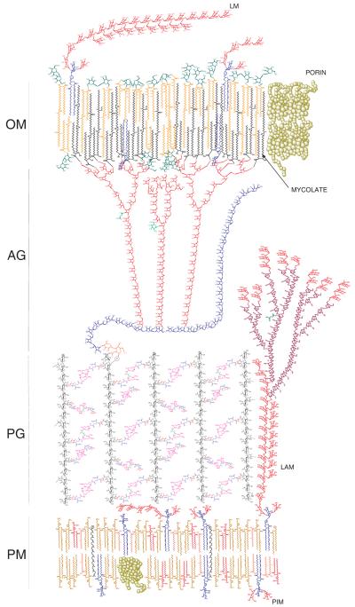
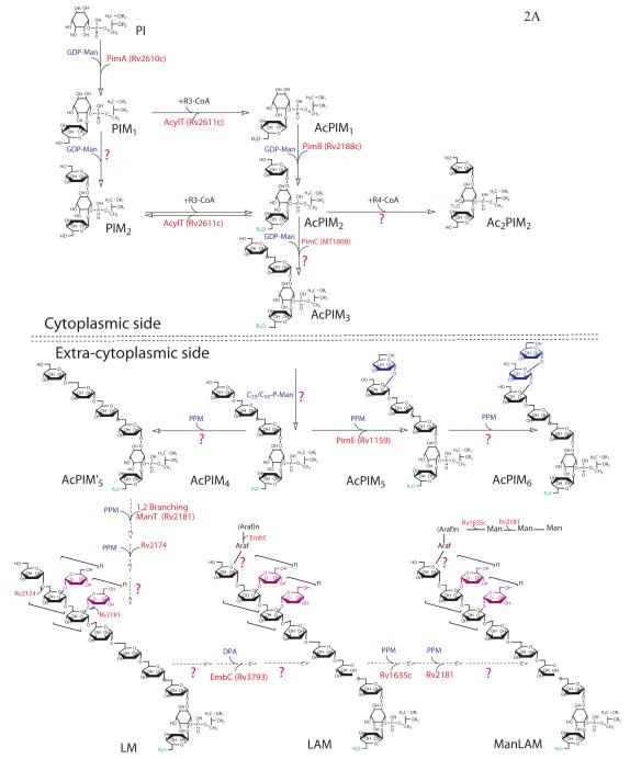
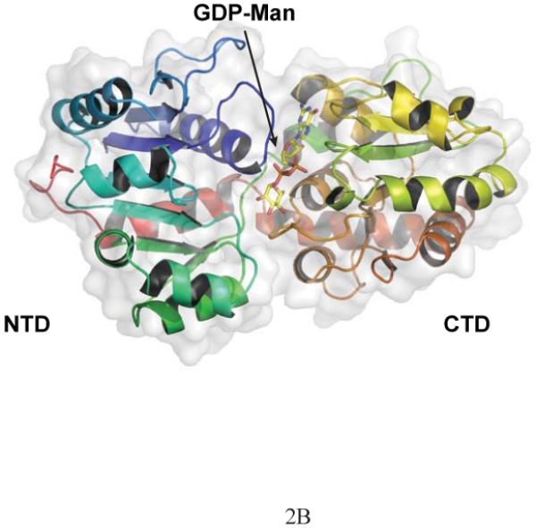
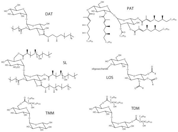
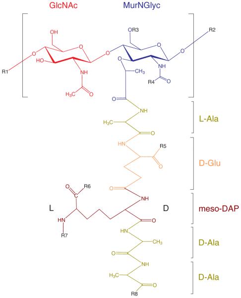
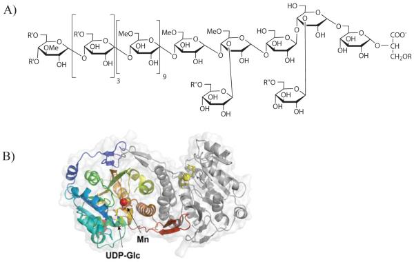
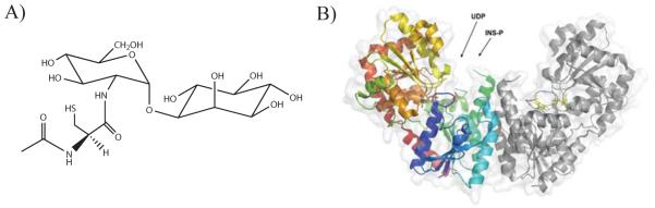
The re-emergence of tuberculosis in its present-day manifestations - single, multiple and extensive drug-resistant forms and as HIV-TB coinfections - has resulted in renewed research on fundamental questions such as the nature of the organism itself, Mycobacterium tuberculosis, the molecular basis of its pathogenesis, definition of the immunological response in animal models and humans, and development of new intervention strategies such as vaccines and drugs. Foremost among these developments has been the precise chemical definition of the complex and distinctive cell wall of M. tuberculosis, elucidation of the relevant pathways and underlying genetics responsible for the synthesis of the hallmark moieties of the tubercle bacillus such as the mycolic acid-arabinogalactan-peptidoglycan complex, the phthiocerol- and trehalose-containing effector lipids, the phosphatidylinositol-containing mannosides, lipomannosides and lipoarabinomannosides, major immunomodulators, and others. In this review, the laboratory personnel who have been the focal point of some to these developments review recent progress towards a comprehensive understanding of the basic physiology and functions of the cell wall of M. tuberculosis.


Figures

Figure 1
Figure 1
A current perspective of the cell envelope of Mycobacterium tuberculosis. Represented is a theoretical model of the outer membrane (OM) wherein similar extractable lipids are present in both leaflets and the meromycolates of bound mycolic acids span the entire hydrophobic region. Another model proposes the meromycolate chains to be folded upon themselves to create a more compact structure, compatible with the observed thickness of the OM (see text for details). The capsular material is not represented here. Extractable lipids represented in the outer membrane include PIMs and acyltrehaloses whereas those of the plasma membrane include phospholipids and PIMs. PM, plasma membrane; PG, peptidoglycan; AG, arabinogalactan; OM, outer membrane. The Galf and Araf residues of AG are represented in blue and red, respectively. The succinyl and galactosamine residues of AG are in green. Manp residues in PIM, LM and LAM are in red; Araf residues are purple.
Figure 2
Figure 2
Biogenesis of PIM, LM and LAM. A) Schematic representation of the current understanding of the PIM, LM and LAM biosynthetic pathways in M. tuberculosis. B) Three-dimensional structure of a PimA-GDP-Man complex. Cartoon representation of the monomeric form of PimA from M. smegmatis. The structure is “color-ramped” from the N-terminus (NTD) (blue) to the C-terminus (CTD) (red).
Figure 2
Figure 2
Biogenesis of PIM, LM and LAM. A) Schematic representation of the current understanding of the PIM, LM and LAM biosynthetic pathways in M. tuberculosis. B) Three-dimensional structure of a PimA-GDP-Man complex. Cartoon representation of the monomeric form of PimA from M. smegmatis. The structure is “color-ramped” from the N-terminus (NTD) (blue) to the C-terminus (CTD) (red).
Figure 3
Figure 3
Structures of some trehalose-derived molecules from Mycobacterium tuberculosis. The major sulfolipid SL-I (2,3,6,6′-tetraacyl α-α’-trehalose-2′-sulfate) is represented. In SL-I, trehalose is sulfated at the 2′ position and esterified with palmitic acid and the multimethyl-branched phthioceranic and hydroxyphthioceranic acids. In DAT (2,3-di-O-acyltrehalose), trehalose is esterified with stearic acid and the multimethyl-branched mycosanoic acid. In PAT (polyacyltrehalose), trehalose is esterified with stearic acid and the multimethyl-branched mycolipenic acids. In TMM and TDM, trehalose is esterified with mycolic acids. The oligosaccharide of the LOS of M. tuberculosis Canetti strains consists of 2-O-methyl-α-L-Fucp-(1→3)-β–D-Glcp-(1→3)-2-O-methyl-α-L-Rhap-(1→3)-2-O-methyl-α-L-Rhap-(1→3)-β-D-Glcp-(1→3)-4-O-methyl-α-L-Rhap-(1→3)-6-O-methyl-α-D-Glc-(1→1)-α-D-Glc. R are 2,4-dimethylhexadecanoic acid and 2,4,6,8-tetramethyloctadecanoic acid residues.
Figure 4
Figure 4
Structures of phenolic glycolipids and p-hydroxybenzoic acid derivatives from M. tuberculosis. The lipid core of PGL from M. tuberculosis is composed of phenolphthiocerol esterified by mycocerosic acids (m = 15-17; n = 20-22, n’, n” = 16, 18; p, p’ = 2-5; R = -CH2-CH3 or –CH3). The trisaccharide substituent of PGL and p-HBAD-II consists of 2,3,4-tri-O-methyl-α-L-Fucp-(1→3)-α–L-Rhap-(1→3)-2-O-methyl-α-L-Rhap. The monosaccharide substituent found in p-HBAD-I consists of 2-O-methyl-α-L-Rhap.
Figure 5
Figure 5
Structure of the predominant mannosyl-β-1-phosphomycoketides from M. tuberculosis H37Rv.
Figure 6
Figure 6
Proposed pathway for the biosynthesis of mycobacterial arabinogalactan. AG synthesis is initiated by a transfer of GlcNAc-1-phosphate onto a polyprenyl-phosphate and continues with the sequential addition of glycosyl residues to this lipid carrier. The enzymes that have been proposed to be involved in this process are indicated on the figure.
Figure 7
Figure 7
A schematic representation of the M. tuberculosis H37Rv cell wall biosynthetic gene cluster (Rv3779-Rv3809c) encompassing genes involved (or likely to be) in the biosynthesis of mycolic acids (accD4, pks13, fadD32, fbpD, fbpA), arabinogalactan (glfT1, Rv3790, Rv3791, atfA, embA, embB, atfB, ubiA, glfT2, glf), and LAM (embA, embB, embC, atfB,, ubiA). Genes annotated or suggested as glycosyltransferases are marked in bold.
Figure 8
Figure 8
Structure of a representative monomer of mycobacterial PG prior to peptide trimming. R1, N-glycolylmuramic acid residue of another monomer; R2, N-acetylglucosamine residue of another monomer; R3, H or the linker unit of AG; R4, H, COCH3 (N-acetyl) or COCH2OH (N-glycolyl); R5, R6, R8, OH, NH2 or OCH3; R7, H, or cross-linked to penultimate d-Ala or to the d-center of another meso-DAP residue.
Figure 9
Figure 9
Methylglucose lipolysaccharides. A) Structure of the MGLPs from M. bovis BCG. The non-reducing end of the polymer is acylated by a combination of acetate, propionate and isobutyrate (R’), whereas octanoate (R) esterifies the position 1 of glyceric acid and zero to three succinate groups (R”) esterify the Glc residues of the reducing end. MGLPs occur as a mixture of four main components that differ in their content of esterified succinic acid. B) Three-dimensional structure of the glucosyl-3-phosphoglycerate synthase from M. avium subsp. paratuberculosis (MAP2569c) in complex with UDP-Glc. Cartoon representation of the dimeric form of the MAP2569c. One monomer is “color-ramped” from the N-terminus (blue) to the C-terminus (red). The second monomer is in grey and UDP-Glc is shown in yellow.
Figure 10
Figure 10
Mycothiol. A) Structure of mycothiol; B) Three-dimensional structure of a CgMshA-UDP-Ins-P complex. Cartoon representation of the dimeric form of CgMshA. One monomer is “color-ramped” from the N-terminus (blue) to the C-terminus (red). The second monomer is in grey and the UDP and Ins-P substrates are shown in yellow. The dimer interface is entirely composed of residues from the N-terminal domain.

References

    1. Adam A, Petit JF, Wietzerbin-Falszpan J, Sinay P, Thomas DW, Lederer E. Mass spectrometric identification of N-glycolylmuramic acid, a constituent of Mycobacterium smegmatis walls. FEBS Lett. 1969;4:87–92. - PubMed
    1. Alderwick LJ, Seidel M, Sahm H, Besra GS, Eggeling L. Identification of a novel arabinosyl transferase (AtfA) involved in cell wall arabinan biosynthesis in Mycobacterium tuberculosis. J. Biol. Chem. 2006;281:15653–15661. - PubMed
    1. Amin AG, Goude R, Shi L, Zhang J, Chatterjee C, Parish T. EmbA is an essential arabinosyltransferase in Mycobacterium tuberculosis. Microbiology. 2008;154:240–248. - PMC - PubMed
    1. Antoine AD, Tepper BS. Characterization of glycogens from mycobacteria. Arch. Biochem. Biophys. 1969;134:207–213. - PubMed
    1. Antoine AD, Tepper BS. Environmental control of glycogen and lipid content of Mycobacterium tuberculosis. J. Bacteriol. 1969;100:538–539. - PMC - PubMed

Publication types

MeSH terms

LinkOut - more resources