Review
. 2009 Dec;13(5-6):503-9.
doi: 10.1016/j.cbpa.2009.07.026. Epub 2009 Sep 2.

Methods for the proteomic identification of protease substrates

Affiliations
Review

Methods for the proteomic identification of protease substrates

Nicholas J Agard et al. Curr Opin Chem Biol. 2009 Dec.

Abstract

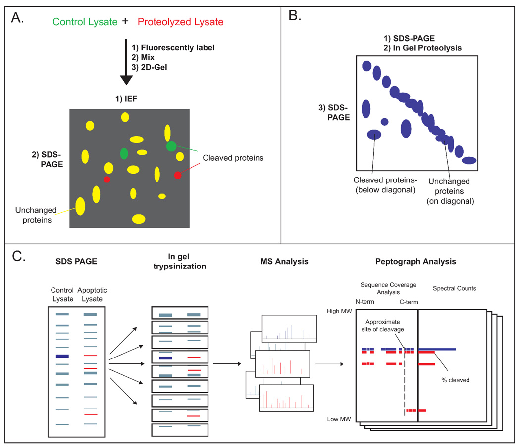
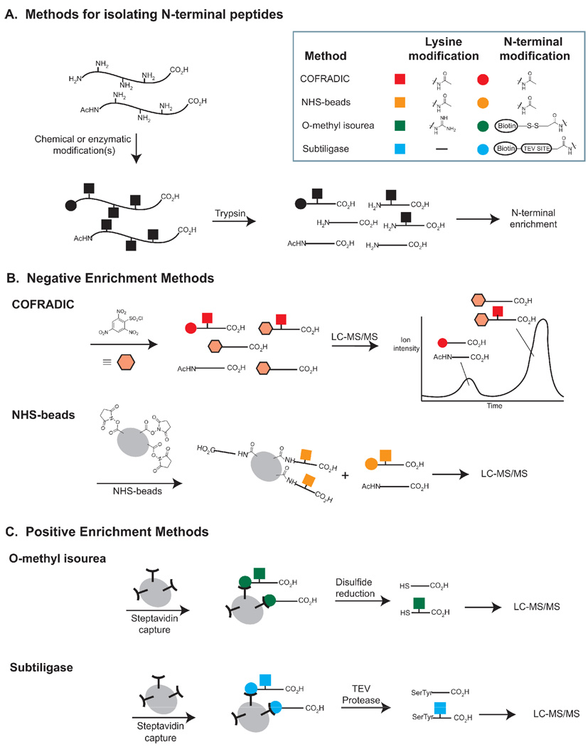
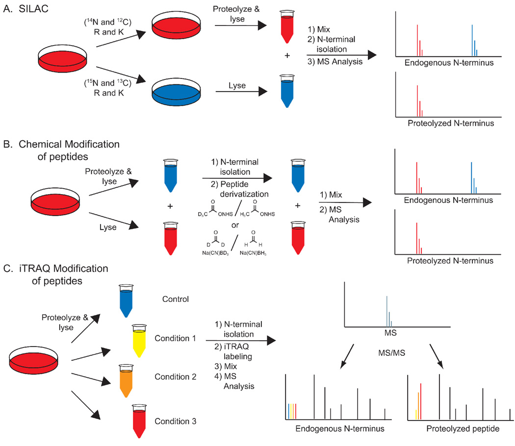
Proteolysis is a key regulatory post-translational modification in diverse cellular processes including programed cell death, immune function, and development. Tracking proteolytic events has become a focus of researchers assessing the downstream consequences of protease activation. In this review we summarize unbiased methods for identifying protease substrates and tracking the extent of cleavage, a field termed 'degradomics'. These include one-dimensional and two-dimensional gel-based methods for identifying protease substrates, N-terminal peptide identification methods for simultaneously identifying substrates and cleavage sites, and approaches for the quantitation of cleavage events during endogenous proteolysis. Individual methods have identified more than 300 caspase-cleaved targets during apoptosis suggesting broad future applications for these technologies.


Figures

Figure 1
Figure 1. Gel-based methods for identifying protease substrates
A. 2-D DIGE: A control and proteolyzed lysate are labeled with fluorescent dyes, mixed, and analyzed by 2-D gel electrophoresis. Spots with unequal fluorescence ratios are picked as proteolyzed proteins for MS analysis. B. 2-D SDS-PAGE: A lysate is resolved by SDS-PAGE, proteolyzed, and electrophoresed perpendicular to the initial migration. Spots occurring below the diagonal are identified as substrates. C. PROTOMAP: Control and proteolyzed cell lysates are analyzed side-by-side via SDS-PAGE. The gel is cut into bands, trypsinized, and peptides are identified by LC-MS/MS. For each protein, peptides are analyzed by peptographs (analysis tools that display the sequence coverage and intensity in each band), revealing the approximate site(s) and extent of cleavage.
Figure 2
Figure 2. Methods for isolating N-terminal Peptides
A. Lysates are treated with chemical reagents or enzymes to modify protein amines. Subsequent trypsinization reveals N-terminal peptides with derivatized N-termini and internal peptides with free amines. B. In negative enrichment methods, internal peptides are removed by reaction with an amine-reactive reagent followed by chromatography or solid phase extraction. The remaining peptides are analyzed by LC-MS/MS. C. In positive enrichment methods, N-terminally labeled peptides are isolated by bead capture, and chemically or enzymatically released for analysis by LC-MS/MS.
Figure 3
Figure 3. Methods for relative peptide quantification
A. SILAC. Duplicate cell cultures are grown in media containing light/heavy amino acids, proteolyzed separately, mixed, and then the N-termini are isolated. Relative peptide intensities are determined by comparing MS intensities. B. MS-based peptide quantification. Cells are lysed, protelyzed, and their N-termini are isolated and derivatized with isotopically labeled reagents. The modified peptides are mixed and quantified based on MS intensities. C. MSMS-based peptide quantification. Isolated N-termini are labeled with isobaric iTRAQ reagents, each with distinct isotopic compositions, and mixed. At the MS level of analysis each peptide is identical. At the MS/MS level the isobaric tag fragments to reveal the relative intensities of the four samples.

References

    1. Muntz K. Protein dynamics and proteolysis in plant vacuoles. J Exp Bot. 2007;58:2391–2407. - PubMed
    1. Doucet A, Butler GS, Rodriguez D, Prudova A, Overall CM. Metadegradomics: toward in vivo quantitative degradomics of proteolytic post-translational modifications of the cancer proteome. Mol Cell Proteomics. 2008;7:1925–1951. - PubMed
    1. Kopan R, Ilagan MX. The canonical Notch signaling pathway: unfolding the activation mechanism. Cell. 2009;137:216–233. - PMC - PubMed
    1. Brasier AR. The NF-kappaB regulatory network. Cardiovasc Toxicol. 2006;6:111–130. - PubMed
    1. Roberts HR, Monroe DM, Hoffman M. Molecular biology and biochemistry of the coagulation factors and pathways of hemostasis. In: Lichtman MA, Kipps JA, Kaushansky K, Beutler E, Seligsohn U, editors. Williams Hematology. edn 7th. Prchal JT: McGraw-Hill; 2006. pp. 1665–1695.

Publication types

Substances

LinkOut - more resources