. 2009 Nov;20(11):2338-47.
doi: 10.1681/ASN.2008111196. Epub 2009 Sep 3.

Embryonic stem cells proliferate and differentiate when seeded into kidney scaffolds

Affiliations

Embryonic stem cells proliferate and differentiate when seeded into kidney scaffolds

Edward A Ross et al. J Am Soc Nephrol. 2009 Nov.

Abstract

The scarcity of transplant allografts for diseased organs has prompted efforts at tissue regeneration using seeded scaffolds, an approach hampered by the enormity of cell types and complex architectures. Our goal was to decellularize intact organs in a manner that retained the matrix signal for differentiating pluripotent cells. We decellularized intact rat kidneys in a manner that preserved the intricate architecture and seeded them with pluripotent murine embryonic stem cells antegrade through the artery or retrograde through the ureter. Primitive precursor cells populated and proliferated within the glomerular, vascular, and tubular structures. Cells lost their embryonic appearance and expressed immunohistochemical markers for differentiation. Cells not in contact with the basement membrane matrix became apoptotic, thereby forming lumens. These observations suggest that the extracellular matrix can direct regeneration of the kidney, and studies using seeded scaffolds may help define differentiation pathways.


Figures
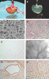
Figure 1.
Figure 1.
Perfusion decellularization and histology of whole rat kidneys. (A) Photograph of harvested kidney with arterial and ureteral cannulae. (B) Photograph of translucent acellular kidney after SDS protocol. (C) Scanning electron micrograph of acellular glomerulus and adjacent tubules showing continuous basement membrane architecture (scale bar, 300 μm) after sodium deoxycholate protocol. Renal cortex before (D, H&E stain) and after (E, H&E; F, bright field) SDS decellularization showing preserved cortical ECM microstructure, with no evidence of residual nuclei or intact cells. Immunohistochemical stains verifying retention of the key basement membrane proteins (G) laminin in the cortical region of the scaffold and (H) collagen IV in the medulla.
Figure 2.
Figure 2.
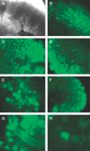
Distribution of green fluorescence protein (GFP)-labeled murine ES cells delivered into the renal artery of decellularized kidney scaffold after (A through G) SDS protocol, thick section culture, and (H) sodium deoxycholate protocol, whole organ perfusion. Images of ES cells at 1 d of culture showing initial glomerular localization in (A) a bright field micrograph (×40) and the corresponding field of view in (B) a fluorescence micrograph (×40). Fluorescence images showing cell growth and migration patterns in cultures up to 6 d. Outgrowth of cells into the interstitium (C) at 3 d (×40); movement of cells into the medulla (D) at 3 d (×40); further expansion of injected cells into a vascular tree (E) at day 4 (×100); and formation of complex branching networks (F) (×40), including larger vessels (G), (×100), at day 6. Fluorescence micrograph showing periglomerular expansion of cells (H) after 6 d using the pulsatile pressure-controlled perfusion device (×150).
Figure 3.
Figure 3.
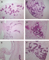
Morphologic patterns of ES cell growth in acellular kidney scaffold, H&E stains, SDS protocol, thick section culture. After antegrade arterial seeding (A through D): cells at day 4 showing uniform localization within glomeruli and adjacent arterioles (A) (×100); cells at day 6 extending into larger vessel lumens and some peritubular capillary networks (B) (×100) and also growing deeper in the medulla (C) (×100); and by day 10 forming a reticular pattern in the cortex interstitium (D) (×400). After retrograde ureteral seeding (E and F): at day 6, cells lining a calyx (E) and achieving cortical distribution (F).
Figure 4.
Figure 4.
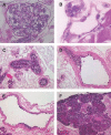
Distinct morphologies of ES cells cultured in the acellular kidney scaffold. After 10 d of whole organ culture (PAS stain, sodium deoxycholate protocol): cells filling the glomerular capillary tufts (A) (×1250) and flat-appearing cells lining vascular structures (B) (×1500). After thick section culture (H&E stain, SDS protocol): at day 6, apoptosis of cells in vessel lumens (C) (×400, a higher magnification of a field in Figure 3B); at day 8, cell aggregates within artery (D) (×400); at day 10, cells lining an artery (E) (×400); and at day 10, solid round cellular aggregates with central apoptosis (F) (×400).
Figure 5.
Figure 5.
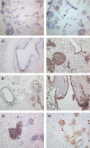
Immunohistochemical analysis of recellularized rat kidneys after SDS protocol and thick section culture. Positive immunoreactivity for pancytokeratin at day 10 in cells within glomeruli and blood vessels (A and B) (×100 and ×200, respectively) and lining arteries (C) (×400). Positive immunoreactivity for Pax-2 at day 6 in glomeruli (D), blood vessels (E), and calyx (F). Positive immunoreactivity for Ksp-cadherin at day 4 in cells in the cortex (G) (×400). Positive immunoreactivity for KI-67 at day 3 in the cortex (H) (×20).
Figure 6.
Figure 6.
Temporal gene expression pattern of Pax-2 and Ksp-cadherin after seeding mES cells in the decellularized scaffold. Total RNA was extracted at the indicated days and subjected to real-time RT-PCR.

References

    1. Lanza R, Langer R, Vicanti J: Principles of Tissue Engineering San Diego, Academic Press, 2000
    1. Atala A: Recent applications of regenerative medicine to urologic structures and related tissues. Curr Opin Urol 16: 305–309, 2006 - PubMed
    1. Gilbert TW, Sellaro TL, Badylak SF: Decellularization of tissues and organs. Biomaterials 27: 3675–3683, 2006 - PubMed
    1. Matsunuma H, Kagami H, Narita Y, Hata K, Ono Y, Ohshima S, Ueda M: Constructing a tissue-engineered ureter using a decellularized matrix with cultured uroepithelial cells and bone marrow-derived mononuclear cells. Tissue Eng 12: 509–518, 2006 - PubMed
    1. Grauss RW, Hazekamp M, Oppenhuizen F, Munsteren C, Gittenberger-de Groot A, DeRuiter M: Histological evaluation of decellularised porcine aortic valves: Matrix changes due to different decellularisation methods. Eur J Cardiothorac Surg 27: 566–571, 2005 - PubMed

Publication types