. 2010 Jan;59(Pt 1):108-114.
doi: 10.1099/jmm.0.012617-0.

Metabolism of azo dyes by human skin microbiota

Affiliations

Metabolism of azo dyes by human skin microbiota

Robin L Stingley et al. J Med Microbiol. 2010 Jan.

Abstract

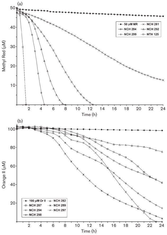
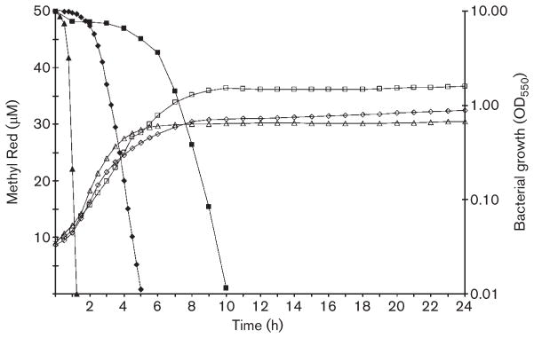
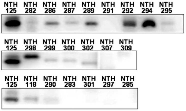
Reduction of Methyl Red (MR) and Orange II (Or II) by 26 human skin bacterial species was monitored by a rapid spectrophotometric assay. The analysis indicated that skin bacteria, representing the genera Staphylococcus, Corynebacterium, Micrococcus, Dermacoccus and Kocuria, were able to reduce MR by 74-100 % in 24 h, with only three species unable to reduce completely the dye in that time. Among the species tested, only Corynebacterium xerosis was unable to reduce Or II to any degree by 24 h, and only Staphylococcus delphini, Staphylococcus sciuri subsp. sciuri and Pseudomonas aeruginosa were able to reduce completely this dye within 24 h. MR reduction started with early-exponential growth in Staphylococcus aureus and Staphylococcus epidermidis, and around late-exponential/early-stationary growth in P. aeruginosa. Reduction of Or II, Ponceau S and Ponceau BS started during late-exponential/early-stationary growth for all three species. Using liquid chromatography/electrospray ionization mass spectrometry analyses, MR metabolites produced by Staph. aureus, Staph. epidermidis and P. aeruginosa were identified as N,N-dimethyl-p-phenylenediamine and 2-aminobenzoic acid. Searches of available genomic and proteomic data revealed that at least four of the staphylococci in this study, Staphylococcus haemolyticus, Staph. epidermidis, Staphylococcus cohnii and Staphylococcus saprophyticus, have hypothetical genes with 77, 76, 75 and 74 % sequence identity to azo1 encoding an azoreductase from Staph. aureus and hypothetical proteins with 82, 80, 72 and 74 % identity to Azo1, respectively. In addition, Staphylococcus capitis has a protein with 79 % identity to Azo1. Western analysis detected proteins similar to Azo1 in all the staphylococci tested, except Staph. delphini, Staph. sciuri subsp. sciuri and Staphylococcus auricularis. The data presented in this report will be useful in the risk assessment process for evaluation of public exposure to products containing these dyes.


Figures

Fig. 1
Fig. 1
Representative dye reduction curves. (a) MR reduction. Among the species that completely reduced 50 μM MR within 24 h, six required 1.5 h or less (represented by Staph. aureus, NTH 125); seven required between 3.5 and 5.5 h (represented by Staphylococcus warneri, NCH 299); four required between 7 and 9 h (represented by Staphylococcus hominis subsp. hominis, NCH 292); and six required between 11 and 14.5 h (represented by M. lylae, NCH 284). Of the 26 bacterial species tested, only three did not completely reduce MR by 24 h (represented by D. nishinomiyaensis, NCH 281). (b) Or II reduction. Only three species were able to completely reduce Or II within 24 h (represented by Staph. sciuri subsp. sciuri, NCH 297). Among the remaining species, no distinct patterns of reduction were observed.
Fig. 2
Fig. 2
Bacterial growth phase during reduction of MR. Reduction of 50 μM MR (filled shapes) was measured at 430 nm and bacterial growth (open shapes) was measured at 550 nm simultaneously. Representative reduction and growth curves are shown for Staph. aureus (NTH 125, triangles), Staph. epidermi-dis (NTH 118, diamonds) and P. aeruginosa (NCH 304, squares). The representative curves shown were from an initial inoculum of OD550 0.05.
Fig. 3
Fig. 3
Western blotting of bacterial protein using a polyclonal anti-Azo1 antiserum. Lanes are labelled using the isolate identification numbers in Table 1. Staph. aureus (NTH 125) protein was used as a positive control for each set of blots. Blots for NCH 281, 284, 303, 304 and 305 are not shown due to non-specific binding of the Azo1 antibody to multiple proteins in these samples.

References

    1. Altschul SF, Madden TL, Schäffer AA, Zhang J, Zhang Z, Miller W, Lipman DJ. Gapped BLAST and PSI-BLAST: a new generation of protein database search programs. Nucleic Acids Res. 1997;25:3389–3402. - PMC - PubMed
    1. Altschul SF, Wootton JC, Gertz EM, Agarwala R, Morgulis A, Schäffer AA, Yu YK. Protein database searches using compositionally adjusted substitution matrices. FEBS J. 2005;272:5101–5109. - PMC - PubMed
    1. Brige A, Motte B, Borloo J, Buysschaert G, Devreese B, Van Beeumen JJ. Bacterial decolorization of textile dyes is an extracellular process requiring a multicomponent electron transfer pathway. Microbial Biotechnol. 2008;1:40–52. - PMC - PubMed
    1. Chen H. Recent advances in azo dye degrading enzyme research. Curr Protein Pept Sci. 2006;7:101–111. - PMC - PubMed
    1. Chen H, Wang RF, Cerniglia CE. Molecular cloning, overexpression, purification, and characterization of an aerobic FMN-dependent azoreductase from Enterococcus faecalis. Protein Expr Purif. 2004;34:302–310. - PMC - PubMed

Publication types

MeSH terms

LinkOut - more resources