

Involvement of sphingosine 1-phosphate (SIP)/S1P3 signaling in cholestasis-induced liver fibrosis
- PMID: 19729475
- PMCID: PMC2751543
- DOI: 10.2353/ajpath.2009.090037
Involvement of sphingosine 1-phosphate (SIP)/S1P3 signaling in cholestasis-induced liver fibrosis
Abstract
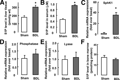
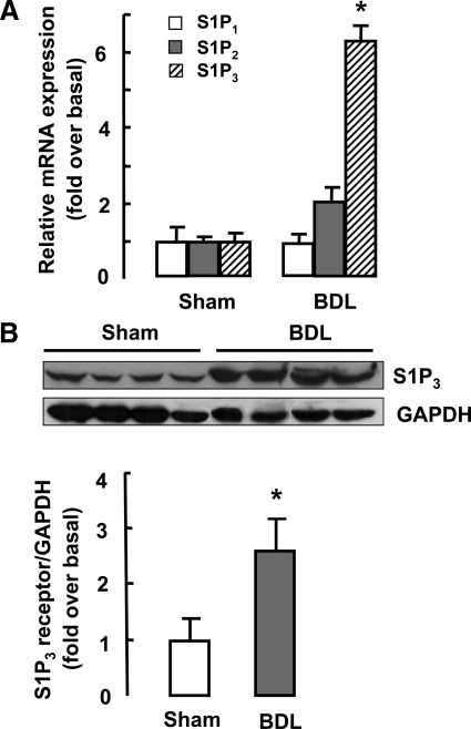
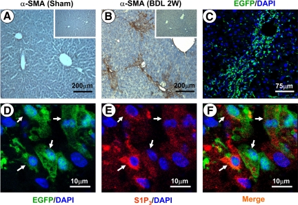
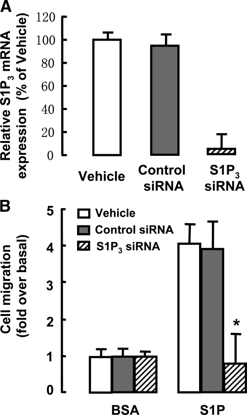
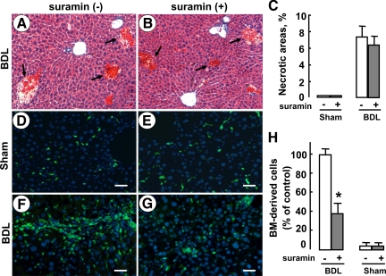
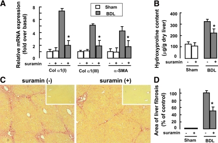
Bioactive sphingosine 1-phosphate (S1P) and S1P receptors (S1PRs) have been implicated in many critical cellular events, including inflammation, cancer, and angiogenesis. However, the role of S1P/S1PR signaling in the pathogenesis of liver fibrosis has not been well documented. In this study, we found that S1P levels and S1P(3) receptor expression in liver tissue were markedly up-regulated in a mouse model of cholestasis-induced liver fibrosis. In addition, the S1P(3) receptor was also expressed in green fluorescent protein transgenic bone marrow (BM)-derived cells found in the damaged liver of transplanted chimeric mice that underwent bile duct ligation. Silencing of S1P(3) expression significantly inhibited S1P-induced BM cell migration in vitro. Furthermore, a selective S1P(3) receptor antagonist, suramin, markedly reduced the number of BM-derived cells during cholestasis. Interestingly, suramin administration clearly ameliorated bile duct ligation-induced hepatic fibrosis, as demonstrated by attenuated deposition of collagen type I and III, reduced smooth muscle alpha-actin expression, and decreased total hydroxyproline content. In conclusion, our data suggest that S1P/S1P(3) signaling plays an important role in cholestasis-induced liver fibrosis through mediating the homing of BM cells. Modulation of S1PR activity may therefore represent a new antifibrotic strategy.
Figures
Similar articles
-
Homing of bone marrow mesenchymal stem cells mediated by sphingosine 1-phosphate contributes to liver fibrosis.J Hepatol. 2009 Jun;50(6):1174-83. doi: 10.1016/j.jhep.2009.01.028. Epub 2009 Mar 29. J Hepatol. 2009. PMID: 19398237
-
The role of sphingosine 1-phosphate receptor 2 in bile-acid-induced cholangiocyte proliferation and cholestasis-induced liver injury in mice.Hepatology. 2017 Jun;65(6):2005-2018. doi: 10.1002/hep.29076. Epub 2017 Apr 28. Hepatology. 2017. PMID: 28120434 Free PMC article.
-
Sphingosine kinase/sphingosine 1-phosphate (S1P)/S1P receptor axis is involved in liver fibrosis-associated angiogenesis.J Hepatol. 2013 Jul;59(1):114-23. doi: 10.1016/j.jhep.2013.02.021. Epub 2013 Mar 4. J Hepatol. 2013. PMID: 23466305
-
Sphingosine-1-phosphate/sphingosine-1-phosphate receptor 1 and T cell migration.Yao Xue Xue Bao. 2016 Jun;51(6):873-8. Yao Xue Xue Bao. 2016. PMID: 29878740 Review.
-
Mechanisms of sphingosine 1-phosphate receptor signalling in cancer.Cell Signal. 2017 Jun;34:66-75. doi: 10.1016/j.cellsig.2017.03.002. Epub 2017 Mar 14. Cell Signal. 2017. PMID: 28302566 Review.
Cited by
-
Expression of sphingosine kinase 1 and sphingosine 1-phosphate receptor 3 in malaria-associated acute lung injury/acute respiratory distress syndrome in a mouse model.PLoS One. 2019 Sep 4;14(9):e0222098. doi: 10.1371/journal.pone.0222098. eCollection 2019. PLoS One. 2019. PMID: 31483837 Free PMC article.
-
Therapeutic targets for cholestatic liver injury.Expert Opin Ther Targets. 2016;20(4):463-75. doi: 10.1517/14728222.2016.1103735. Epub 2015 Oct 19. Expert Opin Ther Targets. 2016. PMID: 26479335 Free PMC article. Review.
-
Sphingosine kinase 1 promotes liver fibrosis by preventing miR-19b-3p-mediated inhibition of CCR2.Hepatology. 2018 Sep;68(3):1070-1086. doi: 10.1002/hep.29885. Epub 2018 Apr 27. Hepatology. 2018. PMID: 29572892 Free PMC article.
-
The beneficial effect of suramin on monocrotaline-induced pulmonary hypertension in rats.PLoS One. 2013 Oct 15;8(10):e77073. doi: 10.1371/journal.pone.0077073. eCollection 2013. PLoS One. 2013. PMID: 24143201 Free PMC article.
-
Divergent Role of Sphingosine 1-Phosphate in Liver Health and Disease.Int J Mol Sci. 2018 Mar 3;19(3):722. doi: 10.3390/ijms19030722. Int J Mol Sci. 2018. PMID: 29510489 Free PMC article. Review.
References
-
- Keeffe EB. Liver transplantation: current status and novel approaches to liver replacement. Gastroenterology. 2001;120:749–762. - PubMed
-
- Kim WR, Ludwig J, Lindor KD. Variant forms of cholestatic diseases involving small bile ducts in adults. Am J Gastroenterol. 2000;95:1130–1138. - PubMed
-
- Alvarez SE, Milstien S, Spiegel S. Autocrine and paracrine roles of sphingosine-1-phosphate. Trends Endocrinol Metab. 2007;18:300–307. - PubMed
Publication types
MeSH terms
Substances
LinkOut - more resources
Full Text Sources
Other Literature Sources
Medical
Molecular Biology Databases
Miscellaneous
