. 2009 Oct;175(4):1746-56.
doi: 10.2353/ajpath.2009.081141. Epub 2009 Sep 3.

Alterations in aortic cellular constituents during thoracic aortic aneurysm development: myofibroblast-mediated vascular remodeling

Affiliations

Alterations in aortic cellular constituents during thoracic aortic aneurysm development: myofibroblast-mediated vascular remodeling

Jeffrey A Jones et al. Am J Pathol. 2009 Oct.

Abstract

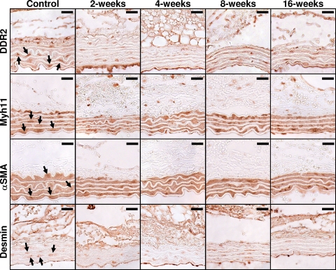
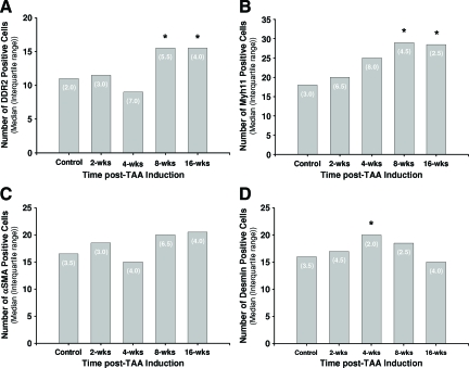
The present study tested the hypothesis that changes in the resident endogenous cellular population accompany alterations in aortic collagen and elastin content during thoracic aortic aneurysm (TAA) development in a murine model. Descending thoracic aortas were analyzed at various time points (2, 4, 8, and 16 weeks) post-TAA induction (0.5 M CaCl2, 15 minutes). Aortic tissue sections were subjected to histological staining and morphometric analysis for collagen and elastin, as well as immunostaining for cell-type-specific markers to quantify fibroblasts, myofibroblasts, and smooth-muscle cells. Results were compared with reference control mice processed in the same fashion. Aortic dilatation was accompanied by changes in the elastic architecture that included: a decreased number of elastic lamellae (from 6 to 4); altered area fraction of elastin (elevated at 4 weeks and decreased at 16 weeks); and a decreased area between elastic lamellae (minimum reached at 4 weeks). Total collagen content did not change over time. Increased immunoreactivity for fibroblast and myofibroblast markers was observed at 8- and 16-week post-TAA-induction, whereas immunoreactivity for smooth-muscle cell markers peaked at 4 weeks and returned to baseline by 16 weeks. Therefore, this study demonstrated that changes in aortic elastin content were accompanied by the emergence of a subset of fibroblast-derived myofibroblasts whose altered phenotype may play a significant role in TAA development through the enhancement of extracellular matrix proteolysis.


Figures

Figure 1
Figure 1
Change in aortic diameter over time post-TAA induction. Median aortic diameter was increased over baseline (average baseline value = 655.3 μm) at each time point post-TAA induction, and diameters in the 2-, 4-, and 8-week groups were different from the 16-week group. Bars represent median aortic diameter; numbers in parentheses are interquartile ranges (25 to 75%). Statistical analysis: Kruskal-Wallis (P = 0.0291); Mann-Whitney (*P < 0.05 versus baseline, **P < 0.05 versus 16-week TAA).
Figure 2
Figure 2
Analysis of medial and adventitial collagen content. Collagen content was measured in aortic tissues from control and TAA-induced mice. Picrosirius red-stained sections were illuminated with polarized light and the resulting birefringence was quantitated in the aortic adventitia (A) and aortic media (B). No significant changes in collagen content were observed. Treatment groups: control (n = 4), 2 weeks (n = 4), 4 weeks (n = 3), 8 weeks (n = 4), and 16 weeks (n = 4). Bars represent median values; numbers in parentheses are interquartile ranges (25 to 75%). Statistical analysis: Kruskal-Wallis (A, P = 0.3696; B, P = 0.3302).
Figure 3
Figure 3
Analysis of medial elastin content and architecture. The medial elastin content and architecture were analyzed in aortic tissues from control and TAA-induced mice. A: Representative Luna-stained sections showing architectural changes over time post-TAA induction; scale bar is equal to 20 μm. B: The number of unbroken elastic lamellae were quantitated at each time point post-TAA induction, Kruskal-Wallis (P = 0.0102). C: The medial area fraction of elastin content was quantitated at each time point post-TAA induction, Kruskal-Wallis (P = 0.0110). D: The gap area between elastic lamellae was quantitated at each time point post-TAA induction, Kruskal-Wallis (P = 0.0136). Treatment groups: control (n = 4), 2 weeks (n = 4), 4 weeks (n = 3), 8 weeks (n = 4), and 16 weeks (n = 4). Bars represent median values; numbers in parentheses are interquartile ranges (25 to 75%). Statistical analysis: Mann-Whitney (*P < 0.05 versus control, **P < 0.05 versus 4-week, and ***P < 0.05 versus 8-week).
Figure 4
Figure 4
Analysis of medial thinning and aortic stiffness over time post-TAA induction. A: Aortic medial thinning was approximated by calculating the ratio of gap area to elastin area, Kruskal-Wallis (P = 0.0087). B: Aortic stiffness was approximated by calculating the ratio of total collagen area to elastin area, Kruskal-Wallis (P = 0.0978). Treatment groups: control (n = 4), 2 weeks (n = 4), 4 weeks (n = 3), 8 weeks (n = 4), and 16 weeks (n = 4). Bars represent median values; numbers in parentheses are interquartile ranges (25 to 75%). Statistical analysis: Mann-Whitney (*P < 0.05 versus control).
Figure 5
Figure 5
Immunohistochemical results for cell-type-specific marker proteins in aortic sections over time post-TAA induction. Aortic tissue sections from and control and TAA-induced mice were stained with cell-type-specific antibodies for fibroblasts (DDR2), SMCs/myofibroblasts (α-SMA and Myh11), and SMCs (desmin). The arrows point to representative cells selected as having positive staining. Color inversion was used to verify cellularity and increased staining intensity over background. Scale bar equals 20 μm.
Figure 6
Figure 6
Quantitation of immunohistochemical staining results for cell-type-specific marker proteins. The median number of positively stained cells in each aortic tissue section from control and TAA-induced mice was determined for each cell-type-specific marker protein: fibroblasts (DDR2), Kruskal-Wallis (P = 0.0122, A); SMCs/myofibroblasts (Myh11), Kruskal-Wallis (P = 0.0216, B); SMCs/myofibroblasts (α-SMA), Kruskal-Wallis (P = 0.2667, C); and SMCs (desmin), Kruskal-Wallis (P = 0.0916, D). Treatment groups: control (n = 4), 2 weeks (n = 4), 4 weeks (n = 3), 8 weeks (n = 4), and 16 weeks (n = 4). Bars represent median values; numbers in parentheses are interquartile ranges (25 to 75%). Statistical analysis: Mann-Whitney (*P < 0.05 versus control).
Figure 7
Figure 7
Immunostaining for apoptotic cells. A: Tissue sections from control and TAA-induced mice were immunostained for the active fragment of caspase-3 (p17) demonstrating enhanced apoptosis at 8- and 16-week post-TAA induction. B: Serial tissue sections from a 16-week TAA were stained with antibodies for apoptotic cells (active caspase-3) or SMCs (desmin). The arrows indicate cells staining for both active caspase-3 and desmin on serial sections. Taken together, these results suggest that a portion of the SMC population undergoes apoptosis during TAA formation.
Figure 8
Figure 8
Summary of cell-type-specific changes over time post-TAA induction. The number of cells staining positive with a specific cell-type marker were expressed as a percent change from control to show relative changes in medial cellular makeup over the course of TAA development. The results demonstrate an increase in the number of DDR2/Myh11/α-SMA+ cells, and a decrease in the number of desmin/α-SMA+ cells, suggesting the emergence of a population of fibroblast-derived myofibroblasts between 4- and 16-week post-TAA induction.

References

    1. Coady MA, Rizzo JA, Goldstein LJ, Elefteriades JA. Natural history, pathogenesis, and etiology of thoracic aortic aneurysms and dissections. Cardiol Clin. 1999;17:615–635. - PubMed
    1. Isselbacher EM. Thoracic and abdominal aortic aneurysms. Circulation. 2005;111:816–828. - PubMed
    1. Barbour JR, Spinale FG, Ikonomidis JS. Proteinase systems and thoracic aortic aneurysm progression. J Surg Res. 2007;139:292–307. - PubMed
    1. Koullias GJ, Ravichandran P, Korkolis DP, Rimm DL, Elefteriades JA. Increased tissue microarray matrix metalloproteinase expression favors proteolysis in thoracic aortic aneurysms and dissections. Ann Thorac Surg. 2004;78:2106–2110. discussion 2110–2101. - PubMed
    1. Dingemans KP, Teeling P, Lagendijk JH, Becker AE. Extracellular matrix of the human aortic media: an ultrastructural histochemical and immunohistochemical study of the adult aortic media. Anat Rec. 2000;258:1–14. - PubMed

Publication types