Comparative Study
. 2009 Oct;175(4):1638-52.
doi: 10.2353/ajpath.2009.090384. Epub 2009 Sep 3.

Comparative analysis of metastasis variants derived from human prostate carcinoma cells: roles in intravasation of VEGF-mediated angiogenesis and uPA-mediated invasion

Affiliations
Comparative Study

Comparative analysis of metastasis variants derived from human prostate carcinoma cells: roles in intravasation of VEGF-mediated angiogenesis and uPA-mediated invasion

Erin M Conn et al. Am J Pathol. 2009 Oct.

Abstract

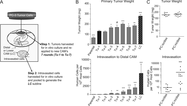
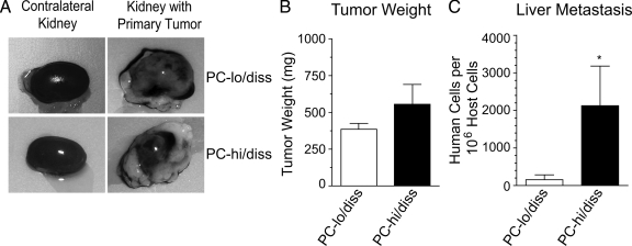
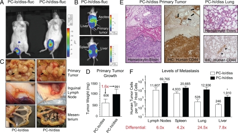
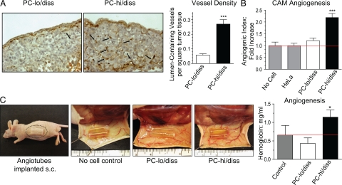
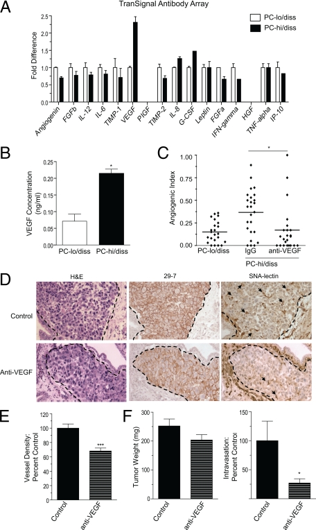
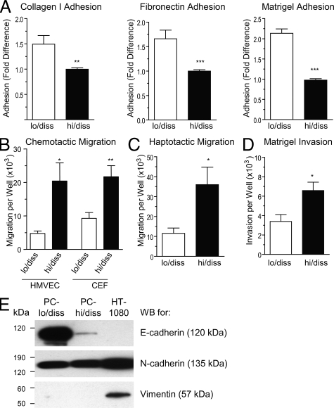
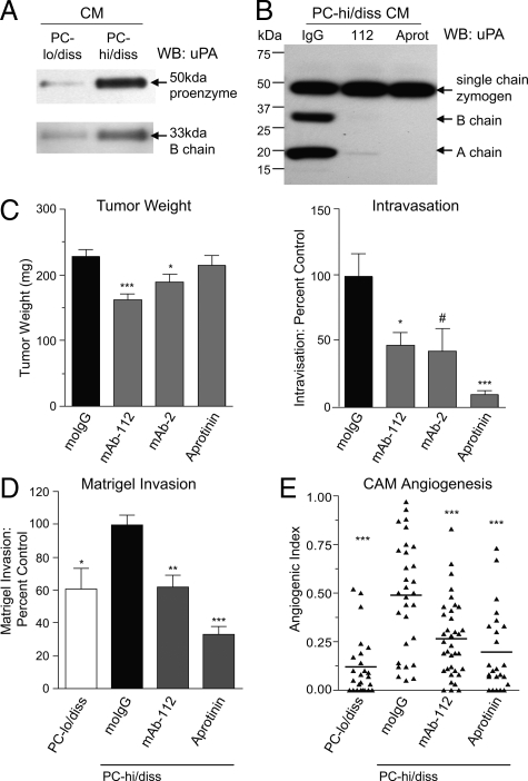
To analyze the process of tumor cell intravasation, we used the human tumor-chick embryo spontaneous metastasis model to select in vivo high (PC-hi/diss) and low (PC-lo/diss) disseminating variants from the human PC-3 prostate carcinoma cell line. These variants dramatically differed in their intravasation and dissemination capacities in both chick embryo and mouse spontaneous metastasis models. Concomitant with enhanced intravasation, PC-hi/diss exhibited increased angiogenic potential in avian and murine models. PC-hi/diss angiogenesis and intravasation were dependent on increased secretion of vascular endothelial growth factor (VEGF), since treating developing tumors with a function-blocking anti-VEGF antibody simultaneously inhibited both processes without affecting primary tumor growth. PC-hi/diss cells were also more migratory and invasive, suggestive of heightened ability to escape from primary tumors due to matrix-degrading activity. Consistent with this suggestion, PC-hi/diss cells produced more of the serine protease urokinase-type plasminogen activator (uPA) as compared with PC-lo/diss. The functional role of uPA in PC-hi/diss dissemination was confirmed by inhibition of invasion, angiogenesis, and intravasation with specific function-blocking antibodies that prevented uPA activation and blocked uPA activity. These processes were similarly sensitive to aprotinin, a potent inhibitor of serine proteases, including uPA-generated plasmin. Thus, our comparison of the PC-3 intravasation variants points to key roles for the uPA-plasmin system in PC-hi/diss intravasation, possibly via (1) promoting tumor cell matrix invasion and (2) facilitating development of VEGF-dependent angiogenic blood vessels.


Figures

Figure 1
Figure 1
In vivo selection of PC-3 dissemination variants and their characterization in the chick embryo spontaneous metastasis model. A: Schematic presentation of multiround selection of dissemination variants from the prostate carcinoma PC-3 cell line. Following grafting on the CAM, the developed tumors were harvested, expanded in vitro and re-applied to new CAMs. A total of 7 rounds of in vitro-in vivo passaging were performed, yielding a series of primary tumor-derived cell lines (Tu-1 to Tu-7). After the seventh round, intravasated cells were isolated from the distal CAM (“Lower CAM”), yielding the LC cell line. B: Isolated PC-3 cell lines were analyzed for their efficiency in tumor growth (top) and intravasation (bottom) in the chick embryo spontaneous metastasis model. Data are means ± SEM from 1 to 5 independent experiments performed with each cell variant. C: A retrospective analysis of tumor cell intravasation (bottom) in individual embryos bearing PC-lo/diss and PC-hi/diss primary tumors of similar size, 160 to 200 mg (top). Statistical significance of the differences was analyzed in comparison with Tu-1 cell line: #P < 0.05, one-tailed t-test; *P < 0.05, **P < 0.01; and ***P < 0.0001 in two-tailed t-test (B, top) or Mann-Whitney test (B, bottom; C).
Figure 2
Figure 2
Dissemination of PC-3 variants in the renal capsule spontaneous metastasis model. A: Selected PC-lo/diss and PC-hi/diss cells were grafted under the kidney capsule of nu/nu mice. Large primary tumors developed on the kidneys and almost completely engulfed them within 4 to 5 weeks post implantation (right panels). For comparison, contralateral kidneys are shown on the left. B: The net weight of individual primary PC-lo/diss and PC-hi/diss tumors was estimated by subtracting the weight of the contralateral kidney. Data are means ± SEM from a representative experiment using 4 mice per variant. C: Metastasis levels in the livers of mice bearing PC-lo/diss (open bars) and PC-hi/diss (closed bars), quantified by Alu-qPCR. *P < 0.05 in one-tailed Mann-Whitney test.
Figure 3
Figure 3
Characterization of PC-3 dissemination variants in an orthotopic spontaneous metastasis model. A: Bioluminescence imaging of PC-lo/diss or PC-hi/diss primary tumors in SCID mice 5 weeks after surgical implantation of firefly luciferase-tagged cells (fluc) into anterior prostates. B: Bioluminescence imaging from side (top) and dorsal (bottom) views of a mouse bearing a PC-hi/diss tumor 5 weeks after implantation. High levels of bioluminescence are associated with the primary tumor, liver, spleen, and abdominal ascites. C: Gross morphology of primary tumors, lymph nodes and mesenterium in sacrificed mice. The tumors in the prostate area are circumscribed by dotted lines (top panels). Arrows point to seminal vesicles, arrowheads point to testes. Significantly enlarged inguinal (subiliac) lymph nodes are indicated by circles (middle panels). The mesenterium in PC-hi/diss-bearing mice is heavily inseminated with macroscopic metastatic nodules (bottom panels). D: Primary tumors were excised and weighed, indicating a 1.6-fold difference in tumor size between the two PC-3 variants. E: H&E staining of a PC-hi/diss tumor (left panel) indicates the presence of a tumor cell mass at the bottom of the section and tumor invasion between prostate glands at the top of the section. A muscular layer in the middle is indicated by dotted lines. The presence of human tumor cells invaded the prostate glandular tissue (arrows) was confirmed by immunohistochemical staining with mAb 29-7, specifically recognizing human CD44 (middle panel). A section of lung tissue from a mouse bearing PC-hi/diss tumor (right panels) stained with H&E (top). Immunohistochemical staining for human CD44 (brown) confirms the presence of metastatic lesion indicated by the arrowhead (bottom). F: Levels of metastasis in mice bearing PC-lo/diss (open bars) and PC-hi/diss (closed bars) tumors were determined by Alu-qPCR in the lymph nodes, spleen, lung, and liver. Bars are means ± SEM from two independent experiments using six mice total per variant. Numbers above bars are mean numbers of humans cells quantified in the organ per 106 murine cells. *P < 0.05 in one-tailed Mann-Whitney test.
Figure 4
Figure 4
Angiogenic potential of PC-3 intravasation variants in chick embryo and mouse models. A: Tumor angiogenesis in the CAM spontaneous metastasis model. PC-lo/diss and PC-hi/diss primary CAM tumors were stained with SNA lectin to highlight endothelial cells (left panels). Black arrows point to lumen-containing blood vessels. Vessel density was determined as a ratio of arbitrary grids with lumen-containing vessels per total number of grids analyzed in digital images of tissue sections taken at ×20 original magnification. From 10 to 20 images per tumor in 3 to 5 individual tumors per variable were analyzed to calculate the means ± SEM presented in the bar graph. B: Angiogenic potential of PC-3 variants in the CAM collagen onplant model. PC-lo/diss and PC-hi/diss cells were incorporated into collagen onplants grafted on the CAM of shell-less chick embryos. The onplants containing collagen alone or non-angiogenic HeLa cells were used as negative controls. The angiogenic index was calculated at 72 hours as the number of grids containing vessels over total number of grids scored in four independent experiments (number of onplants, n = 14, no cells; n = 63, HeLa; n = 45, PC-lo/diss; n = 79, PC-hi/diss). The data are presented as fold difference compared with the angiogenic index of collagen onplants containing HeLa cells. ***P < 0.0001 in two-tailed Student’s t-test. C: Angiogenic potential of PC-3 variants in the mouse angiotube model. Silicon tubes (angiotubes) containing collagen alone (no cell control) or PC-hi/diss or PC-lo/diss tumor cells were implanted under the skin of nu/nu mice (left panel). At 3 weeks, the mice were sacrificed, and the skin flaps with the angiotubes exposed, indicating visually more blood vessels around the PC-hi/diss containing tubes than either control (collagen alone) or PC-lo/diss containing tubes (middle panels). Levels of angiogenesis were estimated by measuring hemoglobin concentration in the contents of individual angiotubes (n = 8 per variable) (bar graph). Bars represent means ± SEM from a representative experiment. *P < 0.05 in two-tailed Student’s t-test.
Figure 5
Figure 5
Increased VEGF production contributes to enhanced angiogenesis and intravasation of PC-hi/diss. A: Analysis of pro- and anti-angiogenic factors produced by PC-lo/diss and PC-hi/diss cells. Serum-free CM from PC-lo/diss and PC-hi/diss cell monolayers was assayed by a TranSignal Angiogenesis Antibody Array, indicating differential in secreted VEGF. B: Levels of VEGF in the CM of PC-hi/diss and PC-lo/diss were quantified by a capture ELISA. C: Contribution of VEGF to PC-hi/diss-induced angiogenesis in the CAM collagen onplant model. A function-blocking antibody to VEGF (R&D) was incorporated at 50 μg/ml into collagen onplants containing PC-hi/diss cells (3 × 10⁴ cells per onplant). Angiogenic index was determined at 72 hours in comparison with onplants containing PC-lo/diss cells. In the scattergram, lines represent means from angiogenic indices determined for each individual onplant in a representative experiment using 5 to 6 embryos per variant (23 to 25 onplants). D: Contribution of VEGF to PC-hi/diss-induced angiogenesis in the chick embryo spontaneous metastasis model. Primary PC-hi/diss CAM tumors were treated topically with the control IgG or function-blocking antibody to VEGF. After 4 days, 3 to 5 control or anti-VEGF-treated tumors were harvested, fixed, and stained with H&E, immunostained with mAb-29-7 against human CD44 to identify tumor cells (brown cell membrane staining), or stained with SNA to highlight the vasculature. Dotted lines delineate the tumor borders. Arrows point to lumen-containing blood vessels. E: Vessel density was calculated after scoring lumen-containing blood vessels in control and anti-VEGF-treated PC-hi/diss tumors in a series of SNA stained tumor sections. Images and vessel density analysis are from a representative experiment, in which three tumors were analyzed per variable with 10 to 20 images analyzed per tumor. F: Contribution of VEGF to PC-hi/diss intravasation in the spontaneous metastasis CAM model. At day 7, control IgG and anti-VEGF-treated PC-hi/diss tumors were harvested and weighed (left panel), and intravasated tumor cells in the distal CAM were quantified by Alu-qPCR (right panel). Tumor weights and intravasation data are pooled values from three individual metastasis experiments with total of 25 and 22 embryos treated with control IgG and anti-VEGF, respectively. #P < 0.05 in one-tailed Mann Whitney test; *P < 0.05, **P < 0.01, and ***P < 0.0001 in two-tailed Student’s t-test.
Figure 6
Figure 6
In vitro characteristics of PC-lo/diss and PC-hi/diss cell variants. A: Adhesion of PC-lo/diss and PC-hi/diss cells to type I collagen, fibronectin, or Matrigel. Data are presented as fold differences of PC-lo/diss adhesion in comparison with adhesion levels of PC-hi/diss, determined from 3 independent experiments performed in triplicate. B: Chemotactic migration of PC-lo/diss and PC-hi/diss cells in Transwells was induced by serum-free CM from CEFs or HMVECs placed into the outer chamber. C: Haptotactic migration of PC-lo/diss and PC-hi/diss cells in Transwells was stimulated by type I collagen coated onto undersurface of the membrane. D: Matrigel invasion of PC-lo/diss and PC-hi/diss cells induced by CM from CEFs placed into the outer chamber. Data are presented as means ± SEM of cells recovered from individual outer chambers following a 48 hour migration or invasion in three independent experiments performed in duplicate. *P < 0.05, **P < 0.01, and ***P < 0.001 in two-tailed Student’s t-test. E: Western blot analysis of PC-lo/diss, PC-hi/diss and HT-1080 cell lysates (20 to 50 μg/lane) for E-cadherin, N-cadherin, and vimentin. Position of molecular weight markers is indicated in kDa on the left.
Figure 7
Figure 7
Functional contribution of uPA to PC-hi/diss intravasation, invasion, and angiogenesis. A: Western blot analysis of uPA production by PC-lo/diss and PC-hi/diss cell variants. SDS-PAGE of CM, normalized to equal numbers of adherent cells, was performed under reducing conditions allowing for detection of single chain pro-uPA (top panel) and B chain of the activated uPA enzyme (bottom panel) by goat anti-uPA antibody. B: Western blot analysis of uPA activation by PC-hi/diss cells. CM was harvested from PC-hi/diss cell monolayers incubated for 48 hours with control IgG, mAb-112, or aprotinin. SDS-PAGE of CM samples was performed under reducing conditions. Single chain uPA zymogen, and B chain and A chain of activated two chain uPA were detected by rabbit anti-uPA antibody recognizing all three uPA species. Position of molecular weight markers is indicated in kDa on the left. C: Contribution of uPA activation and activity to PC-hi/diss intravasation. Developing PC-hi/diss CAM tumors were treated topically with control IgG, mAb-112, mAb-2, or aprotinin. After 7 days, primary tumors were weighed (left graph) and intravasation to the distal CAM was quantified by Alu-qPCR (right graph). Presented are means ± SEM from pooled data of three independent experiments using from 22 to 53 embryos per variable. The levels of intravasation are presented as percentage of intravasation determined in the IgG-treated control. D: Contribution of uPA to Matrigel invasion in Transwells. PC-lo/diss or PC-hi/diss cells were stimulated to invade Matrigel toward CM from CEFs. Where indicated, mAb-112 or aprotinin were added to both inner and outer chambers. Presented are means ± SEM from pooled data of three independent experiments performed in duplicate. Data are calculated as percentage of invasion observed in control IgG-treated PC-hi/diss variant. E: Contribution of uPA to CAM angiogenesis. Collagen onplants containing PC-lo/diss cells mixed with control IgG and PC-hi/diss cells mixed with control IgG, mAb-112, or aprotinin were placed onto the CAMs of shell-less chick embryos. The scattergram depicts angiogenic indices in individual collagen onplants from a representative experiment scored at 72 hours; lines are means. *P < 0.05, **P < 0.005, and ***P < 0.001 in two-tailed Student’s t-test; #P < 0.05 in one-tailed Student’s t-test.

Similar articles

Cited by

References

    1. Naito S, Walker SM, Fidler IJ. In vivo selection of human renal cell carcinoma cells with high metastatic potential in nude mice. Clin Exp Metastasis. 1989;7:381–389. - PubMed
    1. Vezeridis MP, Tzanakakis GN, Meitner PA, Doremus CM, Tibbetts LM, Calabresi P. In vivo selection of a highly metastatic cell line from a human pancreatic carcinoma in the nude mouse. Cancer. 1992;69:2060–2063. - PubMed
    1. Pettaway CA, Pathak S, Greene G, Ramirez E, Wilson MR, Killion JJ, Fidler IJ. Selection of highly metastatic variants of different human prostatic carcinomas using orthotopic implantation in nude mice. Clin Cancer Res. 1996;2:1627–1636. - PubMed
    1. Morikawa K, Walker SM, Jessup JM, Fidler IJ. In vivo selection of highly metastatic cells from surgical specimens of different primary human colon carcinomas implanted into nude mice. Cancer Res. 1988;48:1943–1948. - PubMed
    1. Kang Y, Siegel PM, Shu W, Drobnjak M, Kakonen SM, Cordon-Cardo C, Guise TA, Massague J. A multigenic program mediating breast cancer metastasis to bone. Cancer Cell. 2003;3:537–549. - PubMed

Publication types

Substances