A role for transportin in deposition of TTP to cytoplasmic RNA granules and mRNA decay
- PMID: 19729507
- PMCID: PMC2770677
- DOI: 10.1093/nar/gkp717
A role for transportin in deposition of TTP to cytoplasmic RNA granules and mRNA decay
Abstract
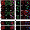
Importin-beta family members, which shuttle between the nucleus and the cytoplasm, are essential for nucleocytoplasmic transport of macromolecules. We attempted to explore whether importin-beta family proteins change their cellular localization in response to environmental change. In this report, we show that transportin (TRN) was minimally detected in cytoplasmic processing bodies (P-bodies) under normal cell conditions but largely translocated to stress granules (SGs) in stressed cells. Fluorescence recovery after photobleaching analysis indicated that TRN moves rapidly in and out of cytoplasmic granules. Depletion of TRN greatly enhanced P-body formation but did not affect the number or size of SGs, suggesting that TRN or its cargo(es) participates in cellular function of P-bodies. Accordingly, TRN associated with tristetraprolin (TTP) and its AU-rich element (ARE)-containing mRNA substrates. Depletion of TRN increased the number of P-bodies and stabilized ARE-containing mRNAs, as observed with knockdown of the 5'-3' exonuclease Xrn1. Moreover, depletion of TRN retained TTP in P-bodies and meanwhile reduced the fraction of mobile TTP to SGs. Therefore, our data together suggest that TRN plays a role in trafficking of TTP between the cytoplasmic granules and whereby modulates the stability of ARE-containing mRNAs.
Figures


Similar articles
-
Protor-2 interacts with tristetraprolin to regulate mRNA stability during stress.Cell Signal. 2012 Jan;24(1):309-15. doi: 10.1016/j.cellsig.2011.09.015. Epub 2011 Sep 22. Cell Signal. 2012. PMID: 21964062 Free PMC article.
-
The mRNA-capping enzyme localizes to stress granules in the cytoplasm and maintains cap homeostasis of target mRNAs.J Cell Sci. 2024 Jun 1;137(11):jcs261578. doi: 10.1242/jcs.261578. Epub 2024 Jun 6. J Cell Sci. 2024. PMID: 38841902
-
TTP and BRF proteins nucleate processing body formation to silence mRNAs with AU-rich elements.Genes Dev. 2007 Mar 15;21(6):719-35. doi: 10.1101/gad.1494707. Genes Dev. 2007. PMID: 17369404 Free PMC article.
-
Stress granules: sites of mRNA triage that regulate mRNA stability and translatability.Biochem Soc Trans. 2002 Nov;30(Pt 6):963-9. doi: 10.1042/bst0300963. Biochem Soc Trans. 2002. PMID: 12440955 Review.
-
Tristetraprolin: A cytosolic regulator of mRNA turnover moonlighting as transcriptional corepressor of gene expression.Mol Genet Metab. 2021 Jun;133(2):137-147. doi: 10.1016/j.ymgme.2021.03.015. Epub 2021 Mar 26. Mol Genet Metab. 2021. PMID: 33795191 Review.
Cited by
-
Identification of potential cargo proteins of transportin protein AtTRN1 in Arabidopsis thaliana.Plant Cell Rep. 2016 Mar;35(3):629-40. doi: 10.1007/s00299-015-1908-4. Epub 2015 Dec 9. Plant Cell Rep. 2016. PMID: 26650834
-
RNA granules: the good, the bad and the ugly.Cell Signal. 2011 Feb;23(2):324-34. doi: 10.1016/j.cellsig.2010.08.011. Epub 2010 Sep 8. Cell Signal. 2011. PMID: 20813183 Free PMC article. Review.
-
Monocyte chemotactic protein-induced protein 1 (MCPIP1) suppresses stress granule formation and determines apoptosis under stress.J Biol Chem. 2011 Dec 2;286(48):41692-41700. doi: 10.1074/jbc.M111.276006. Epub 2011 Oct 4. J Biol Chem. 2011. PMID: 21971051 Free PMC article.
-
A novel PSMB8 isoform associated with multiple sclerosis lesions induces P-body formation.Front Cell Neurosci. 2024 May 15;18:1379261. doi: 10.3389/fncel.2024.1379261. eCollection 2024. Front Cell Neurosci. 2024. PMID: 38812791 Free PMC article.
-
Identification of Novel Stress Granule Components That Are Involved in Nuclear Transport.PLoS One. 2013 Jun 27;8(6):e68356. doi: 10.1371/journal.pone.0068356. Print 2013. PLoS One. 2013. PMID: 23826389 Free PMC article.
References
-
- Gorlich D, Kutay U. Transport between the cell nucleus and the cytoplasm. Annu. Rev. Cell Dev. Biol. 1999;15:607–660. - PubMed
-
- Kuersten S, Ohno M, Mattaj IW. Nucleocytoplasmic transport: ran, beta and beyond. Trends Cell Biol. 2001;11:497–503. - PubMed
-
- Pollard VW, Michael WM, Nakielny S, Siomi MC, Wang F, Dreyfuss G. A novel receptor-mediated nuclear protein import pathway. Cell. 1996;86:985–994. - PubMed
-
- Lai MC, Lin RI, Huang SY, Tsai CW, Tarn WY. A human importin-beta family protein, transportin-SR2, interacts with the phosphorylated RS domain of SR proteins. J. Biol. Chem. 2000;275:7950–7957. - PubMed
Publication types
MeSH terms
Substances
LinkOut - more resources
Full Text Sources
Molecular Biology Databases

