. 2009 Sep 4;325(5945):1230-4.
doi: 10.1126/science.1176811.

A sulfilimine bond identified in collagen IV

Affiliations

A sulfilimine bond identified in collagen IV

Roberto Vanacore et al. Science. .

Abstract

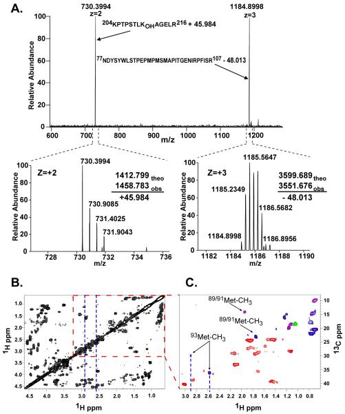
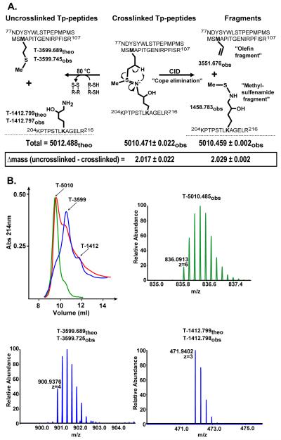
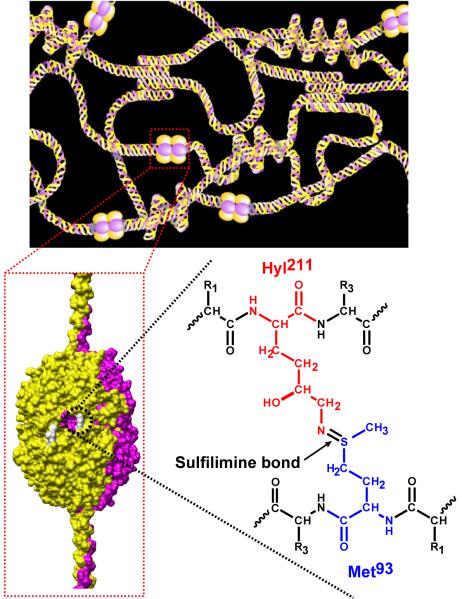
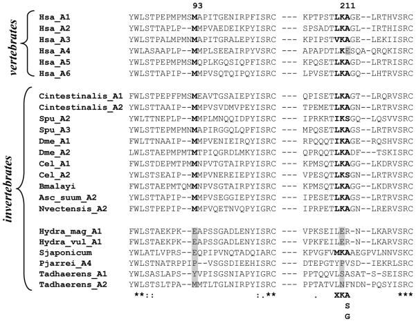
Collagen IV networks are ancient proteins of basement membranes that underlie epithelia in metazoa from sponge to human. The networks provide structural integrity to tissues and serve as ligands for integrin cell-surface receptors. They are assembled by oligomerization of triple-helical protomers and are covalently crosslinked, a key reinforcement that stabilizes networks. We used Fourier-transform ion cyclotron resonance mass spectrometry and nuclear magnetic resonance spectroscopy to show that a sulfilimine bond (-S=N-) crosslinks hydroxylysine-211 and methionine-93 of adjoining protomers, a bond not previously found in biomolecules. This bond, the nitrogen analog of a sulfoxide, appears to have arisen at the divergence of sponge and cnidaria, an adaptation of the extracellular matrix in response to mechanical stress in metazoan evolution.


Figures

Fig. 1
Fig. 1
Mass spectrometric and NMR analyses of the cross-linked Tp-peptides from the α1NC1-α1NC1 dimer. A) MS2 spectrum showing the fragmentation by collision-induced dissociation of the m/z 1003.1014 (+5) ion (Table 1). The bottom panel shows the isotopic envelope for each ion and the mass difference of each fragment with respect to the uncross-linked peptides. B) NMR studies showing the 1H-1H correlated spectroscopy COSY spectrum in 50 mM phosphate buffer, pH=7.0, 20°C. C) Edited 1H-13C HSQC spectrum of an expanded portion of panel B in which the methyl groups of Val, Leu, Thr, Iso, and Met are expected. Correlation peaks are color-coded according to multiplicity edited HSQC spectra, optimized for correlation selection of CH2 (red), CH/CH3 (blue) and CH (green) only. Several peaks contain overlapping signals from CH3 and CH groups are indicated in purple.
Fig. 2
Fig. 2
Summary of the MS analyses of the crosslinked Tp-peptides before and after reduction with DTT. A) The uncross-linked tryptic peptides, T-3599.689 and T-1412.799, derived from the α1-NC1 domain, display the side chains of Met93 and Hyl211, respectively. T-5012.488 corresponds to the total theoretical mass of both peptides. The sulfilimine double bond crosslinking the tryptic peptides is shown. The difference between the theoretical (theo) and observed (obs) mass reveals that two hydrogen atoms are lost upon sHM crosslink formation. Fragmentation of the sulfilimine bond by CID produces peptide fragments containing an olefin fragment derived from Met93 and a methylsulfenamide fragment derived from Hyl211 as a result of the “Cope elimination” event in the gas phase. However, chemical reduction with DTT, formally involving addition of 2 H-atoms, severs the sulfilimine link and recovers Met93 and Hyl211, as indicated below. B) Cross-linked Tp-peptides were separated by gel filtration chromatography before (green) and after incubation in 100 mM DTT at room temperature (red) and 80 °C (blue). The arrows indicate the identity of each chromatographic peak as revealed by MS analysis.
Fig. 3
Fig. 3
Proposed chemical structure of the sHM cross-link. Schematic of the α1α2α1 collagen IV network (top panel) illustrating the interaction between the NC1 domain of triple-helical protomers. A space-filling model of the NC1 hexamer quaternary structure (bottom left) shows the location of the sHM cross-links (white). The sulfilimine bond that constitutes the sHM crosslink connecting the side chains of Met93 and Hyl211 is shown for the α1NC1-α1NC1 dimer. The same bond connects Met93 and Hyl211 in the α2NC1-α2NC1 dimer.
Fig. 4
Fig. 4
Multiple sequence alignment of collagen IV NC1 domain sequences encompassing Met93 and Lys211. Met93 and Lys211 are shown in bold. Conserved amino acid residues are indicated with an asterisk (*). Semi-conserved residues are indicated with a colon (:). The hydroxylation motif for lysyl hydroxylase, X-K-(A/S/G) is shown at the bottom of the alignment. Abbreviations are as follow: Homo sapiens (Hsa; A1, NP_001836.2; A2, NM_001846.2; A3, ABX71213.1; A4, X81053.1; A5, ABW24668.1; A6, AL136080.6), Ciona intestinalis (Cintestinalis, A1, XP_2120982.1; A2, XP_2119477.1), Strongylocentrotus purpuratus (Spu, A2, NP_999676.1; A3, NP_999631.1), Drosophila melanogaster (Dme, A1, AAA28404.1; A2, AAB64082.1), Caenorhabditis elegans (Cel, A1, AAB59179.1; A2, AAA27989.1), Brugia malayi (Bmalayi, XP_1902932.1), Ascaris Suum (Asc_suum, AAA18014.1), Nematostella vectensis (Nvectensis, XP_1626265.1), Hydra magnipapillata (Hydra_mag, XP_2157001.1), Hydra vulgaris (Hydra_vul, AAG40729.1), Schistosoma japonicum (Sjaponcum, AAX25734.2), Trichoplax adhaerens (Tadhaerens, A1, EDV21329.1; A2, EDV21231.1). The alignments were generated with ClustalW.

References

    1. Moser M, Legate KR, Zent R, Fassler R. Science. 2009 May 15;324:895. - PubMed
    1. Hynes RO. Cell. 2002 Sep;110:673. - PubMed
    1. Yurchenco PD, Furthmayr H. Biochemistry. 1984 Apr 10;23:1839. - PubMed
    1. Fox MA, et al. Cell. 2007 Apr 6;129:179. - PubMed
    1. Gould DB, et al. N Engl J Med. 2006 Apr 6;354:1489. - PubMed

Publication types