. 2009 Sep 4;325(5945):1261-5.
doi: 10.1126/science.1173569.

Activation of the PI3K pathway in cancer through inhibition of PTEN by exchange factor P-REX2a

Affiliations

Activation of the PI3K pathway in cancer through inhibition of PTEN by exchange factor P-REX2a

Barry Fine et al. Science. .

Abstract

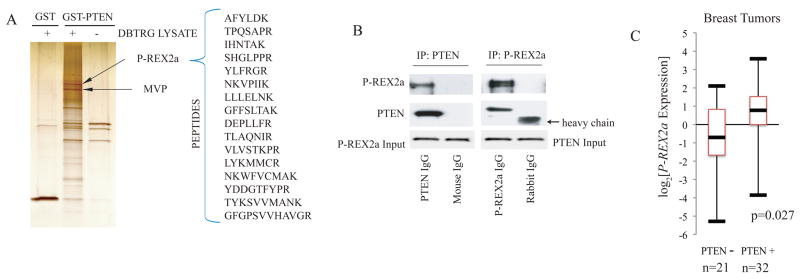
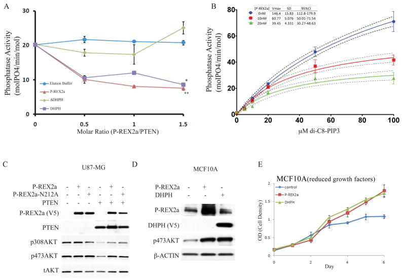
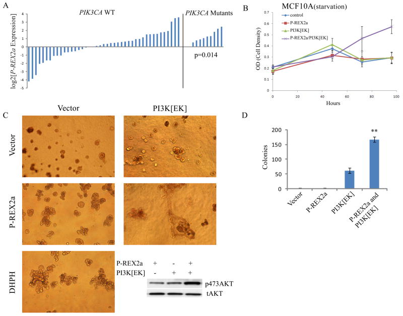
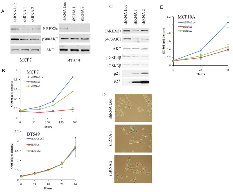
PTEN (phosphatase and tensin homolog on chromosome 10) is a tumor suppressor whose cellular regulation remains incompletely understood. We identified phosphatidylinositol 3,4,5-trisphosphate RAC exchanger 2a (P-REX2a) as a PTEN-interacting protein. P-REX2a mRNA was more abundant in human cancer cells and significantly increased in tumors with wild-type PTEN that expressed an activated mutant of PIK3CA encoding the p110 subunit of phosphoinositide 3-kinase subunit alpha (PI3Kalpha). P-REX2a inhibited PTEN lipid phosphatase activity and stimulated the PI3K pathway only in the presence of PTEN. P-REX2a stimulated cell growth and cooperated with a PIK3CA mutant to promote growth factor-independent proliferation and transformation. Depletion of P-REX2a reduced amounts of phosphorylated AKT and growth in human cell lines with intact PTEN. Thus, P-REX2a is a component of the PI3K pathway that can antagonize PTEN in cancer cells.


Figures

Fig. 1
Fig. 1. P-REX2a as a PTEN-binding protein
(A) Silver stain of 1M salt elutions of affinity purified PTEN binding proteins. GST and GST-PTEN columns were incubated with (left, middle) and without (right) DBTRG-05MG cytoplasmic extract and proteins eluted with high salt were separated and identified by mass spectrometry. P-REX2a and MVP are indicated with arrows. (B) Co-immunoprecipitation of endogenous PTEN and P-REX2a. Immunoprecipitions were performed as per Materials and Methods and proteins were detected by immunoblotting. (C) Box plot of P-REX2a expression in PTEN positive and negative breast tumors. Upper and lower bars represent the maximum and minimum expression respectively. The box delineates the first to third quartiles of expression and the central bar represents the median. P-REX2a levels are significantly associated with PTEN status (p=0.027) by two-tailed t-test.
Fig. 2
Fig. 2. Inhibition of PTEN phosphatase activity by P-REX2a
(A) Full-length P-REX2a, or a deletion of the DHPH domain (ΔDHPH) or the DHPH domain alone were added in the indicated molar volumes to 40nM PTEN, purified from HEK293 cells, and phosphatase activity of PTEN was assayed with 20μM di-C8-PIP3. This is a representative experiment, error bars indicate standard deviation (n=3), **p<0.005, *p<0.05 by ANOVA. (B) P-REX2a (10nM or 20nM) was added to the reactions and phosphate released was measured over a range of di-C8-PIP3 using 40nM PTEN. Regression line (solid) to Michaelis-Menten kinetics is shown along with 95% confidence interval (dotted lines). Vmax values are shown in table along with standard deviation and 95% confidence interval. Representative experiment shown and error bars represent +/− standard deviation (n=3). (C) Effect of P-REX2a and a GEF dead point mutant of P-REX2a, N212A, on phosphorylation of AKT in presence and absence of PTEN. (D) Effect of expression of P-REX2a alone in MCF10A cells on abundance of p473AKT. The phosphorylation status of T308AKT was not detectable under normal growth conditions in MCF10A cells. (E) Effect of P-REX2a and DHPH on proliferation of MCF10A cells grown in reduced growth factors (0.1% serum).
Fig. 3
Fig. 3. P-REX2a and PI3K mutants cooperate
(A) P-REX2a expression levels in PTEN wild-type breast tumors without PIK3CA PI3K activating mutations (right) (p=0.014; two tailed t-test). (B) Proliferation in the absence of all growth factors and serum in MCF10A cells expressing P-REX2a and the PI3K mutant E545K (PI3K[EK]). (C) Microscopy of branched multilobulated mammospheres in MCF10A cells expressing P-REX2a or DHPH as indicated when grown in matrigel. Amount of pAKT shown (inset). All phase contrast images are displayed at 100x magnification. (D) Colony formation by MCF10A cells grown in soft agar. Cells expressing P-REX2a and PI3K[EK] as indicated (**p<0.01; t-test).
Fig. 4
Fig. 4. Diminished phosphorylation of AKT and proliferation after depletion of P-REX2a
Phosphorylation of AKT (A) and proliferation (B) after depletion of P-REX2a in PTEN wild type MCF7 cells or PTEN-null BT549 cells. (C) Effect of P-REX2a depletion in MCF10A cells on pAKT and pGSK3β and amounts of the p21 and p27 cell cycle inhibitors. (D) X-gal staining of indicated cell lines. Phase contrast images displayed at 40x magnification. (E) MCF10A proliferation after P-REX2a depletion.

References

    1. Eng C. Hum Mutat. 2003;22:183. - PubMed
    1. Maehama T, Dixon JE. J Biol Chem. 1998;273:13375. - PubMed
    1. Cantley LC. Science. 2002;296:1655. - PubMed
    1. Cully M, You H, Levine AJ, Mak TW. Nat Rev Cancer. 2006;6:184. - PubMed
    1. Baker SJ. Cell. 2007;128:25. - PubMed

Publication types

MeSH terms

Substances