. 2009 Dec 15;180(12):1179-88.
doi: 10.1164/rccm.200904-0552OC. Epub 2009 Sep 3.

Lung dendritic cell expression of maturation molecules increases with worsening chronic obstructive pulmonary disease

Affiliations

Lung dendritic cell expression of maturation molecules increases with worsening chronic obstructive pulmonary disease

Christine M Freeman et al. Am J Respir Crit Care Med. .

Abstract

Rationale: Dendritic cells (DCs) have not been well studied in chronic obstructive pulmonary disease (COPD), yet their integral role in activating and differentiating T cells makes them potential participants in COPD pathogenesis.

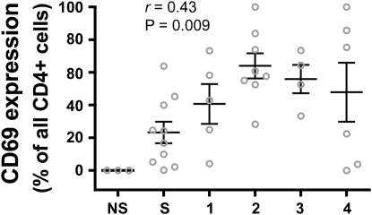
Objectives: To determine the expression of maturation molecules by individual DC subsets in relationship to COPD stage and to expression of the acute activation marker CD69 by lung CD4(+) T cells.

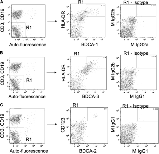
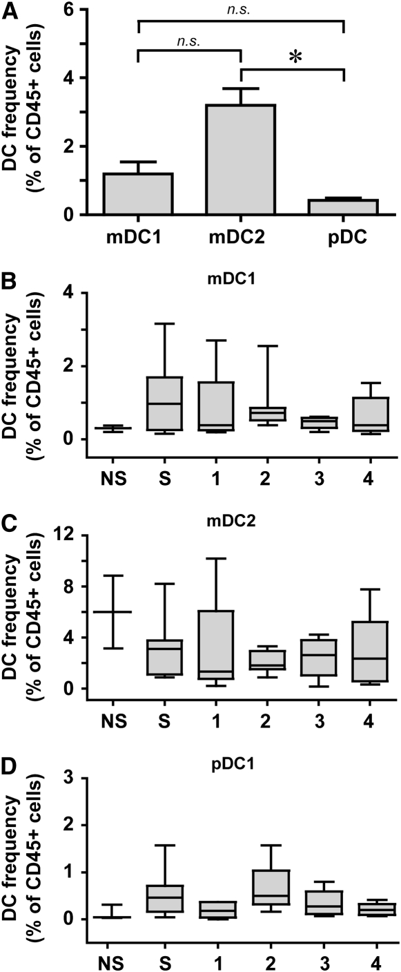
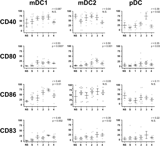
Methods: We nonenzymatically released lung leukocytes from human surgical specimens (n = 42) and used flow cytometry to identify three DC subsets (mDC1, mDC2, and pDC) and to measure their expression of three costimulatory molecules (CD40, CD80 and CD86) and of CD83, the definitive marker of DC maturation. Spearman nonparametric correlation analysis was used to identify significant correlations between expression of DC maturation molecules and COPD severity.

Measurements and main results: Expression of CD40 by mDC1 and mDC2 and of CD86 by mDC2 was high regardless of GOLD stage, but CD80 and CD83 on these two DC subsets increased with disease progression. pDC also showed significant increases in expression of CD40 and CD80. Expression of all but one of the DC molecules that increased with COPD severity also correlated with CD69 expression on lung CD4(+) T cells from the same patients, with the exception of CD83 on mDC2.

Conclusions: This cross-sectional study implies that COPD progression is associated with significant increases in costimulatory molecule expression by multiple lung DC subsets. Interactions with lung DCs may contribute to the immunophenotype of CD4(+) T cells in advanced COPD. Clinical trial registered with www.clinicaltrials.gov (NCT00281229).


Figures

Figure 1.
Figure 1.
Gating strategy used to identify dendritic cell (DC) subsets among human lung leukocytes. Lung tissue was dispersed nonenzymatically and was stained with monoclonal antibodies to identify DC subsets. After gating on CD45+ cells (left column), cells that had low autofluorescence and that were CD3 and CD19 were selected (gate R1). From within gate R1, we then selected cells (middle column) that were (A) BDCA-1+ and HLA-DR+ (mDC1), (B) BDCA-3+ and HLA-DR+ (mDC2), or (C) BDCA-2+ and CD123+ (pDC). Isotype controls are shown in the right column. Representative data are from individual subjects.
Figure 2.
Figure 2.
Myeloid DCs are more prevalent in human lungs than plasmacytoid DCs. (A) Frequency of DC subsets in entire group (n = 42), determined as a percent of CD45+ cells. Data represent the mean ± SEM. * P < 0.001 (one-way analysis of variance, with Dunn's post hoc testing). mDC1 = myeloid dendritic cell type 1; mDC2 = myeloid dendritic cell type 2; n.s.,= nonsignificant; pDC = plasmacytoid dendritic cell. (BD) Frequency of DC subsets stratified by group. (B) mDC1. (C) mDC2. (D) pDC. NS = neversmokers; S = smokers without COPD; 1–4, GOLD stages. Box and whisker plots show the median (central bar), interquartiles (boxes), and range (whiskers) of the data; note differences in scales. There were no significant differences between groups (nonparametric Kruskal-Wallis and Mann-Whitney U tests).
Figure 3.
Figure 3.
All three DC subsets can be identified in lung interstitium in chronic obstructive pulmonary disease (COPD). Frozen tissue sections were stained with antibody against (A) BDCA1 (mDC1), (C) BDCA-3 (mDC2), (E) BDCA-2 (pDC), or (B, D, F) the respective isotype control antibodies. The immunoperoxidase reaction was visualized using 3-amino-9-ethylcarbazole substrate (red) with hematoxylin counterstaining. Magnification: 1,000×, scale = 50 μm.
Figure 4.
Figure 4.
Activation markers on DC subsets increase in correlation with disease severity. Using flow cytometry, expression of CD40, CD80, CD86, and CD83 on mDC1 (left column), mDC2 (middle column), and pDC (right column) subsets was determined and correlated to GOLD stage. The frequency (as percentage of positive cells) of a given marker is shown on the vertical axis, and subject group is shown on the horizontal axis, NS = never-smokers; S = smokers without COPD. Spearman nonparametric correlation analysis generated an r value that was used to determine significance. Open circles represent individual patients. Bars represent the mean ± SEM.
Figure 5.
Figure 5.
CD69 expression on lung CD4+ T cells correlates with COPD severity. Surface expression of CD69 on lung CD4+ T cells was determined by flow cytometry and is expressed as the percentage of all lung CD4+ T cells on the vertical axis; subject groups are shown on the horizontal axis. NS = never-smokers; S = smokers without COPD. Spearman nonparametric correlation analysis is shown. Open circles represent individual patients. Bars represent the mean ± SEM.
Figure 6.
Figure 6.
Lung mDC2 interact physically with lung CD4+ T cells. Frozen lung tissue sections were stained with antibodies against (A) BDCA-3 (immunoperoxidase/3-amino-9-ethylcarbazole, red) and CD4 (alkaline phosphatases, black) or (B) isotype control. Magnification: 1,000×; scale = 50 μm.

Comment in

References

    1. O'Donnell R, Breen D, Wilson S, Djukanovic R. Inflammatory cells in the airways in COPD. Thorax 2006;61:448–454. - PMC - PubMed
    1. Hogg JC, Chu F, Utokaparch S, Woods R, Elliott WM, Buzatu L, Cherniack RM, Rogers RM, Sciurba FC, Coxson HO, et al. The nature of small-airway obstruction in chronic obstructive pulmonary disease. N Engl J Med 2004;350:2645–2653. - PubMed
    1. Lambrecht BN, Hammad H. Taking our breath away: dendritic cells in the pathogenesis of asthma. Nat Rev Immunol 2003;3:994–1003. - PubMed
    1. Freeman CM, Curtis JL, Chensue SW. CC chemokine receptor 5 and CXC chemokine receptor 6 expression by lung CD8+ cells correlates with chronic obstructive pulmonary disease severity. Am J Pathol 2007;171:767–776. - PMC - PubMed
    1. Demedts IK, Bracke KR, Van Pottelberge G, Testelmans D, Verleden GM, Vermassen FE, Joos GF, Brusselle GG. Accumulation of dendritic cells and increased CCL20 levels in the airways of patients with chronic obstructive pulmonary disease. Am J Respir Crit Care Med 2007;175:998–1005. - PubMed

Publication types

Associated data