. 2009 Oct;119(10):3089-101.
doi: 10.1172/JCI37978. Epub 2009 Sep 1.

Cyclin I activates Cdk5 and regulates expression of Bcl-2 and Bcl-XL in postmitotic mouse cells

Affiliations

Cyclin I activates Cdk5 and regulates expression of Bcl-2 and Bcl-XL in postmitotic mouse cells

Paul T Brinkkoetter et al. J Clin Invest. 2009 Oct.

Abstract

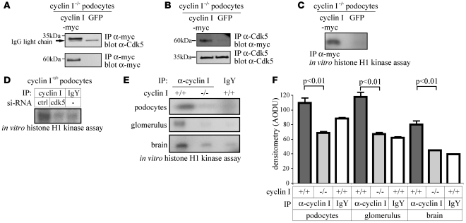
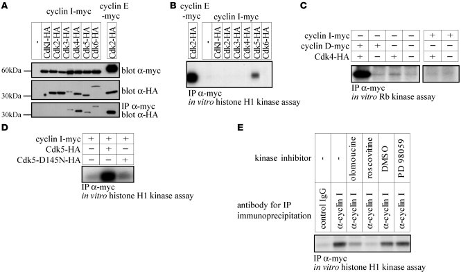
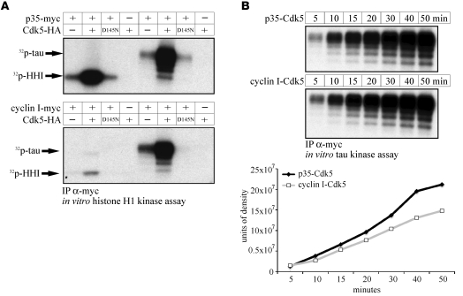
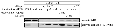
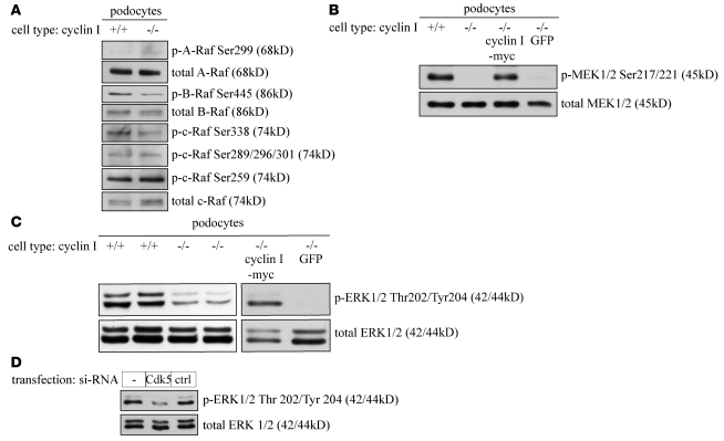
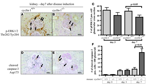
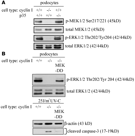
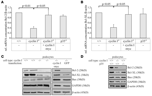
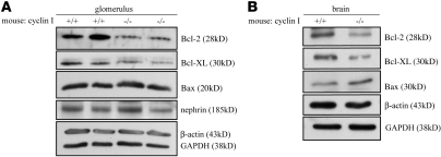
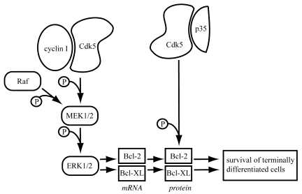
Cyclin I is an atypical cyclin because it is most abundant in postmitotic cells. We previously showed that cyclin I does not regulate proliferation, but rather controls survival of podocytes, terminally differentiated epithelial cells that are essential for the structural and functional integrity of kidney glomeruli. Here, we investigated the mechanism by which cyclin I safeguards against apoptosis and found that cyclin I bound and activated cyclin-dependent kinase 5 (Cdk5) in isolated mouse podocytes and neurons. Cdk5 activity was reduced in glomeruli and brain lysates from cyclin I-deficient mice, and inhibition of Cdk5 increased in vitro the susceptibility to apoptosis in response to cellular damage. In addition, levels of the prosurvival proteins Bcl-2 and Bcl-XL were reduced in podocytes and neurons from cyclin I-deficient mice, and restoration of Bcl-2 or Bcl-XL expression prevented injury-induced apoptosis. Furthermore, we found that levels of phosphorylated MEK1/2 and ERK1/2 were decreased in cyclin I-deficient podocytes and that inhibition of MEK1/2 restored Bcl2 and Bcl-XL protein levels. Of interest, this pathway was also defective in mice with experimental glomerulonephritis. Taken together, these data suggest that a cyclin I-Cdk5 complex forms a critical antiapoptotic factor in terminally differentiated cells that functions via MAPK signaling to modulate levels of the prosurvival proteins Bcl-2 and Bcl-XL.


Figures

Figure 1
Figure 1. Cyclin I binds and activates constitutive Cdk5 in postmitotic cells.
(A, B) To determine whether cyclin I and Cdk5 coimmunoprecipitate in glomerular podocytes, cyclin I–null (–/–) podocytes were infected with either myc-tagged cyclin I or GFP. Reciprocal coimmunoprecipitation studies with either α-myc (A, lanes 1, 2) or α-Cdk5 (B, lanes 3, 4) antibodies revealed that cyclin I bound to endogenous Cdk5 in podocytes. (C) Histone H1 kinase activity was abundant in cyclin I–null podocytes infected with cyclin I–myc (lane 1). In contrast, kinase activity was not detected in cultured cyclin I–null podocytes infected with GFP (lane 2). (D) To prove that the cyclin I–associated histone H1 kinase activity shown in C was specifically due to the activation of Cdk5, WT (+/+) podocytes were transfected with siRNA. Cyclin I–associated histone H1 kinase activity was present in podocytes transfected with control siRNA (lane 1). In contrast, cyclin I–associated kinase activity was not detected in cells transfected with siRNA targeting Cdk5 (lane 2). As expected, an IP with a control preimmune IgY antibody showed no kinase activity (lane 3). Ctrl, control. (E and F) To determine whether cyclin I–Cdk5 was also active in tissues in vivo, kinase activity was measured in protein extracts from kidney glomeruli and brain of WT and cyclin I–null mice. Cdk5 was active in WT cultured podocytes, glomeruli, and brain tissue. In contrast, histone H1 kinase activity was not detected in corresponding tissues from cyclin I–null mice. Densitometric analysis (n = 3). Data shown represent mean + SD.
Figure 2
Figure 2. Cyclin I is a specific binding partner and activator of Cdk5 in HEK293 cells.
(A) Following cotransfection of HEK293 cells with myc-tagged cyclin I and HA-tagged Cdks1–6, cyclin I coimmunoprecipitated with Cdk 3–6. (B, C) To assess which Cdk was activated by cyclin I, immunoprecipitations were performed followed by histone H1 (B) or Rb (C) kinase assays. Kinase activity was only detected in cells cotransfected with cyclin I and Cdk5. HA-Cdk2 and myc–cyclin E (B, histone H1 assay) and HA-Cdk4 and myc–cyclin D (C, Rb assay) served as positive controls. These results show that cyclin I specifically activates Cdk5, but not Cdk1, -2, -3, -4, or -6. (D) To prove specificity, HEK293 cells were cotransfected with cyclin I alone, cyclin I and Cdk5, or cyclin I and a dominant negative Cdk5 (D145N-Cdk5). No kinase activity was detected when cells were transfected with cyclin I alone (lane 1). Kinase activity was detected in cells transfected with cyclin I and Cdk5 (lane 2), but not in cells with cyclin I and the kinase-inactive mutant (lane 3). (E) No histone H1 kinase activity was detected when control IgG was used for immunoprecipitation (lane 1). Following immunoprecipitation with an antibody to myc, kinase activity was present (lane 2). In the presence of the Cdk inhibitors olomoucine (10 μM; lane 3) and roscovitine (50 μM, lane 4), kinase activity was reduced. The controls DMSO and PD98059 (an ERK5 inhibitor) had no effect on cyclin I kinase activity.
Figure 3
Figure 3. Cyclin I–Cdk5 preferentially phosphorylates tau.
(A) HEK293 cells were transfected with HA-Cdk5, then cotransfected with either myc-p35 or myc–cyclin I. Following an IP to the myc epitope tag on either p35 or cyclin I, in vitro kinase assays were performed using either histone H1 (HH1) or tau as substrates. Histone H1 and tau (lanes 2, 6) were strongly phosphorylated by p35-Cdk5. In contrast, cyclin I–Cdk5 preferentially phosphorylated tau (lane 6) rather than histone H1 (lane 2). No kinase activity was detected in the presence of a kinase-inactive dominant negative Cdk5 mutant (HA-D145N-Cdk5, lane 3, 7) or in the absence of either Cdk5 (lanes 1, 5) or p35/cyclin I (lanes 4, 8). (B) To compare the kinase kinetics by which cyclin I–Cdk5 phosphorylates tau in comparison with p35-Cdk5, a time-course experiment was performed in HEK293 cells cotransfected with myc-p35 and HA-Cdk5 or myc–cyclin I and HA-Cdk5. Following IP for the myc tag, phosphorylation of tau was assessed by incorporation of 32P-ATP quantified on a phosphoimager system. Cyclin I–Cdk5 showed phosphorylation kinetics similar to those of p35-Cdk5. Phosphorylation of tau was already evident after 5 minutes and reached a plateau after 40–50 minutes.
Figure 4
Figure 4. Cyclin I–Cdk5 kinase activity is required to protect podocytes from apoptosis.
(A) Apoptosis was quantitated by Hoechst 33342 staining in podocytes under nonstressed conditions (baseline) and 6 hours after UV-C irradiation (25 J/m2). UV-C–induced apoptosis was increased 2- to 3-fold in WT podocytes transfected with siRNA-targeting Cdk5 (lane 3) compared with cells transfected with control siRNA (lane 2). UV-C induced marked apoptosis in cyclin I–null podocytes (lane 4). Experiments were performed in triplicate, and a minimum of 400 cells were counted per experiment. pos, positive. (B) In the absence of UV-C, Cdk5 protein levels measured by Western blot analysis were similar in nontransfected cyclin I WT and null podocytes (lanes 1, 2). siRNA reduced Cdk5 protein levels in WT cells (middle panel), which resulted in increased caspase-3 cleavage following UV-C irradiation (lower panel); caspase-3 cleavage was not increased in control siRNA–transfected cells exposed to this dose of UV-C (lanes 6, 7). β-actin served as loading control. (C) UV-C irradiation increased apoptosis (Hoechst 33342 staining) 3- to 4-fold in cyclin I WT podocytes when Cdk5 activity was inhibited by roscovitine (lane 3) compared with control cells exposed to DMSO (lane 2). (D) Roscovitine induced a progressive increase in cleaved caspase-3 (lanes 5, 6) following exposure to UV-C. β-actin served as a loading control. Data shown represent mean + SD.
Figure 5
Figure 5. Reducing p35 or Cdk5 activity augments caspase-3 cleavage in cyclin I–null podocytes.
To determine the relative roles of cyclin I, p35, and Cdk5 on apoptosis measured by caspase-3 cleavage, cyclin I–null podocytes were transfected (tx) with siRNA targeting either Cdk5 or p35, and in separate studies, Cdk5 activity was reduced in cyclin I–null cells with roscovitine. Reducing Cdk5 protein levels augmented caspase-3 cleavage in cyclin I–null cells following UV-C irradiation compared with control siRNA–transfected cells (lanes 1, 2). Lowering p35 protein levels increased caspase-3 cleavage following UV-C irradiation in cyclin I–null cells compared with nontransfected and control siRNA–transfected cells (lanes 3–5). Roscovitine increased caspase-3 cleavage (lane 6) following exposure to UV-C to the levels seen in cyclin I–null cells transfected with siRNA that targets p35.
Figure 6
Figure 6. Cyclin I–Cdk5 regulates apoptosis by activating MEK1/2–ERK1/2.
(A) There were no substantial differences in the phosphorylation status of A-, B- and c-Raf in cyclin I–null (–/–) and WT (+/+) podocytes under nonstressed conditions using several phosphospecific antibodies. (B) Phosphorylation of MEK1/2 on residues Ser217/221 was substantially reduced in cyclin I–null podocytes (lane 2) compared with WT podocytes (lane 1) under physiological, nonstressed conditions. Restoring cyclin I levels in null cells by retroviral infection normalized MEK1/2 phosphorylation (lane 3) to levels comparable to those of WT podocytes. GFP transfection had no effect (lane 4). Total MEK served as a loading control. These results show that MEK1/2 Ser217/221 phosphorylation is cyclin I dependent. (C) Phosphorylation of ERK1/2 on residues Thr202/Tyr204 was reduced in 2 different clones of cyclin I–null podocytes (lanes 3, 4) compared with 2 different WT podocyte clones (lanes 1, 2). Restoring cyclin I levels in null cells by retroviral infection normalized ERK1/2 phosphorylation (lane 5) to levels comparable to those of WT podocytes; GFP infection had no effect. Total ERK1/2 served as loading control. These results show that the cyclin I–dependent activation of MEK1/2 was reflected by an increased phosphorylation of ERK1/2. (D) Reducing Cdk5 expression in cyclin I WT podocytes with siRNA decreased ERK1/2 phosphorylation (lane 2) compared with nontransfected cells. Scrambled siRNA had no effect on ERK1/2 phosphorylation.
Figure 7
Figure 7. Decreased ERK1/2 activation and increased caspase-3 cleavage in cyclin I–null mice with experimental glomerulonephritis.
Experimental glomerulonephritis was induced in 10- to 12-week-old WT and cyclin I–null mice by administration of anti-glomerular antibody. (AC) Activation of ERK1/2 was assessed by immunostaining for p-ERK1/2 Thr202/Tyr204. There was a significant decrease in glomerular pERK1/2 staining at day 7 of nephritis in cyclin I–null mice (P < 0.05, ANOVA). (DF) Apoptosis was quantified by immunostaining for caspase-3 cleavage adjacent to Asp175. Both WT and cyclin I–null mice showed increased caspase-3 cleavage in podocytes following disease induction. However, there was significantly more caspase-3 cleavage in cyclin I–null mice at day 7 compared with WT mice (P < 0.01, ANOVA). Depicted are representative glomeruli. Data shown represent mean + SD.
Figure 8
Figure 8. The signaling pathway downstream of cyclin I–Cdk5 is distinct from siRNA that targets p35-Cdk5.
(A) There were no differences in MEK1/2 and ERK1/2 phosphorylation in the absence of p35 in podocytes compared with WT podocytes. (B) To prove that the specific decrease in MEK1 was central to the increases in apoptosis in the absence of cyclin I, cyclin I–null podocytes were transfected with constitutively active MEK1 (MEK-DD) mutants. Restoring active MEK1 increased ERK 1/2 phosphorylation (lane 3) similar to that of WT cells (lane 1). Restoring MEK1 reduced UV-C–induced apoptosis measured by caspase-3 cleavage products. These results show that cyclin I is required to phosphorylate MEK1 (and thus ERK1/2), which is necessary to reduce apoptosis.
Figure 9
Figure 9. Cyclin I, but not p35, differentially regulates specific Bcl-2 family proteins.
(A, B) The mRNA levels for Bcl-2 (A) and Bcl-XL (B) were measured by quantitative PCR, and the relative concentrations are shown as ratio normalized to β-actin. Samples were run in triplicate, and mRNA from 3 independent experiments was included. Relative gene expression was analyzed using the 2–standard curve method. Compared with WT cells (lanes 1), the mRNA levels for Bcl-2 and Bcl-XL (lanes 2) were significantly reduced in cyclin I–null podocytes. Restoring cyclin I in null cells by stable infection normalized transcripts for Bcl-2 and Bcl-XL (lanes 3). In contrast, the absence of p35 in p35-null cells had no effect on Bcl-2 and Bcl-XL mRNA levels (lane 4). (C, D) To determine whether cyclin I or p35 altered the protein levels for certain Bcl-2 family proteins, Western blot analyses were performed in WT, cyclin I–null, and p35-null podocytes grown under physiological, nonstressed conditions. GAPDH and β-actin served as loading controls. Compared with WT cells (C, lanes 1, 2), Bcl-2 and Bcl-XL, but not Bax, protein levels were reduced in cyclin I–null podocytes (C, lanes 3, 4). Levels for Bcl-2 and Bcl-XL were normalized upon infection with cyclin I (lane 5), but not GFP (lane 6). In contrast, in the absence of p35, only Bcl-2 protein expression was strongly reduced compared with WT podocytes (D). No effects on Bcl-XL and Bax protein levels were observed. Data shown represent mean + SD.
Figure 10
Figure 10. Cyclin I regulates specific Bcl-2 family proteins in vivo.
(A) Glomeruli were isolated and pooled from the kidneys of 6 animals from each strain, divided into 2 samples, and loaded separately on the gel. Compared with WT glomeruli (lanes 1, 2), levels of Bcl-2 and Bcl-XL were reduced in cyclin null glomeruli (lanes 3, 4). Nephrin, a protein expressed selectively and constitutively in podocytes, was included as a loading control in addition to β-actin and GAPDH to rule out any potential loading differences in protein from podocytes. (B) Compared with brain protein lysates harvested from cyclin I WT mice (lane 1) under physiological, nonstressed conditions, the protein levels of Bcl-2 and Bcl-XL were decreased in cyclin I–null mice (lane 2). Bax levels remain unchanged; β-actin was used as loading control. Taken together, these data show that similar to the cultured cells, the absence of cyclin I also regulates Bcl-2 and Bcl-XL in postmitotic organs in vivo.
Figure 11
Figure 11. Cyclin I, but not p35, differentially regulates specific Bcl-2 family proteins.
(A) Reducing Cdk5 expression by siRNA decreased the protein levels of Bcl-2 and Bcl-XL. No effect was observed in control podocytes transfected with control siRNA. (B) Inhibiting Cdk5 activity by roscovitine (50 μM) reduced the protein expression of Bcl-2 and Bcl-XL compared with vehicle (DMSO). GAPDH and β-actin served as loading controls. (C) To prove that regulation of Bcl-2 and Bcl-XL underlies the prosurvival effect of cyclin I, protein expression of Bcl-2 or Bcl-XL was restored in cyclin I–null podocytes by retroviral infection. There was a 3- to 4-fold increase in apoptosis following UV-C irradiation in cyclin I–null cultured podocytes infected with GFP compared with injured cyclin I WT cells. UV-C–induced apoptosis was markedly reduced in cyclin I–null podocytes stably infected with Bcl-2 or Bcl-XL. (D) Accordingly, caspase-3 cleavage products were also decreased in cyclin I–null podocytes infected with Bcl-2 or Bcl-XL 6 hours after UV-C irradiation compared with GFP-infected podocytes. (E) Infecting cyclin I–null podocytes with constitutively active MEK1 (MEK-DD) mutants also increased the expression of Bcl-2 and Bcl-XL linking regulation of the MEK-ERK pathway by cyclin I–Cdk5 to the observed regulation of Bcl-2 and Bcl-XL expression. Data shown represent mean + SD.
Figure 12
Figure 12. Proposed model showing the effects of Cdk5 upon activation by cyclin I and p35.
Cdk5 is activated by cyclin I and p35. The pathways by which Cdk5 confers a prosurvival function depend on the activator. Cyclin I–Cdk5 leads to phosphorylation of MEK1/2, and subsequently, ERK1/2, leading to increased mRNA and protein levels for the prosurvival proteins Bcl-2 and Bcl-XL. In contrast, p35-Cdk5 increases Bcl-2 protein levels by posttranslational modification (30). p35-Cdk5 has no effect on Bcl-XL levels. The dual activation of Cdk5 by cyclin I and p35 ensures that maximal survival pathways are operative in terminally differentiated cells.

References

    1. Kobayashi N., et al. Process formation of the renal glomerular podocyte: is there common molecular machinery for processes of podocytes and neurons? Anat. Sci. Int. 2004;79:1–10. doi: 10.1111/j.1447-073x.2004.00066.x. - DOI - PubMed
    1. Bredesen D.E., Rao R.V., Mehlen P. Cell death in the nervous system. Nature. 2006;443:796–802. doi: 10.1038/nature05293. - DOI - PMC - PubMed
    1. Cho B.B., Toledo-Pereyra L.H. Caspase-independent programmed cell death following ischemic stroke. J. Invest. Surg. 2008;21:141–147. doi: 10.1080/08941930802029945. - DOI - PubMed
    1. Barisoni L., Kriz W., Mundel P., D’Agati V. The dysregulated podocyte phenotype: a novel concept in the pathogenesis of collapsing idiopathic focal segmental glomerulosclerosis and HIV-associated nephropathy. J. Am. Soc. Nephrol. 1999;10:51–61. - PubMed
    1. Pagtalunan M.E., et al. Podocyte loss and progressive glomerular injury in type II diabetes. J. Clin. Invest. 1997;99:342–348. doi: 10.1172/JCI119163. - DOI - PMC - PubMed

Publication types

MeSH terms