. 2009 Sep;5(9):e1000629.
doi: 10.1371/journal.pgen.1000629. Epub 2009 Sep 4.

Establishment of cohesion at the pericentromere by the Ctf19 kinetochore subcomplex and the replication fork-associated factor, Csm3

Affiliations

Establishment of cohesion at the pericentromere by the Ctf19 kinetochore subcomplex and the replication fork-associated factor, Csm3

Josefin Fernius et al. PLoS Genet. 2009 Sep.

Abstract

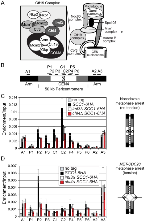
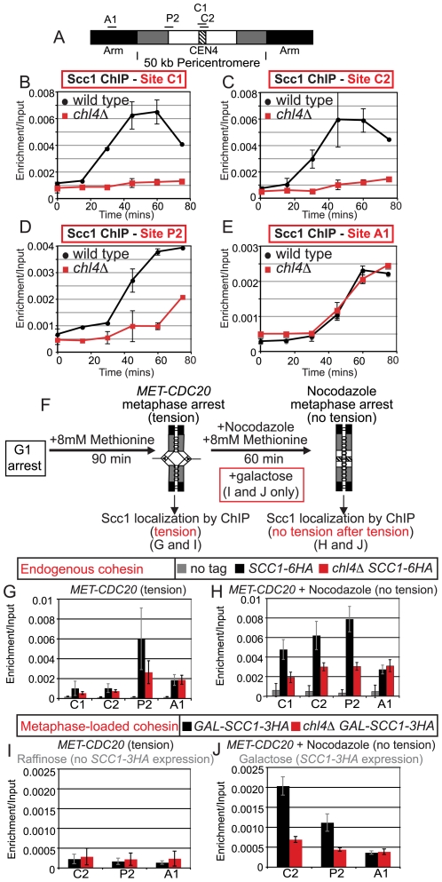
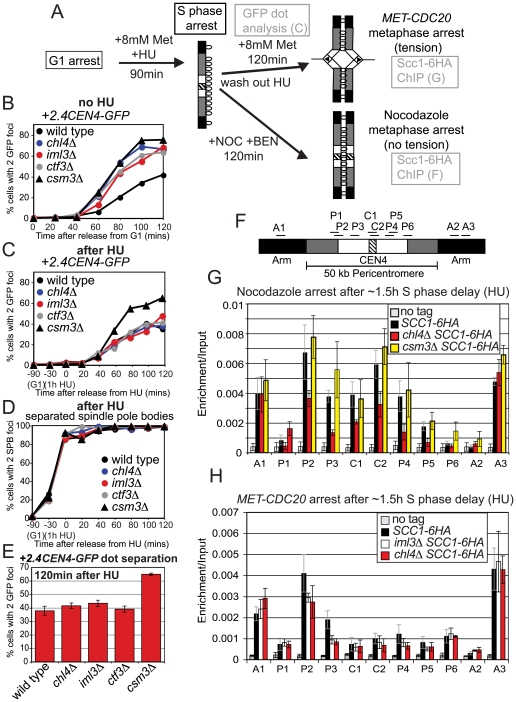
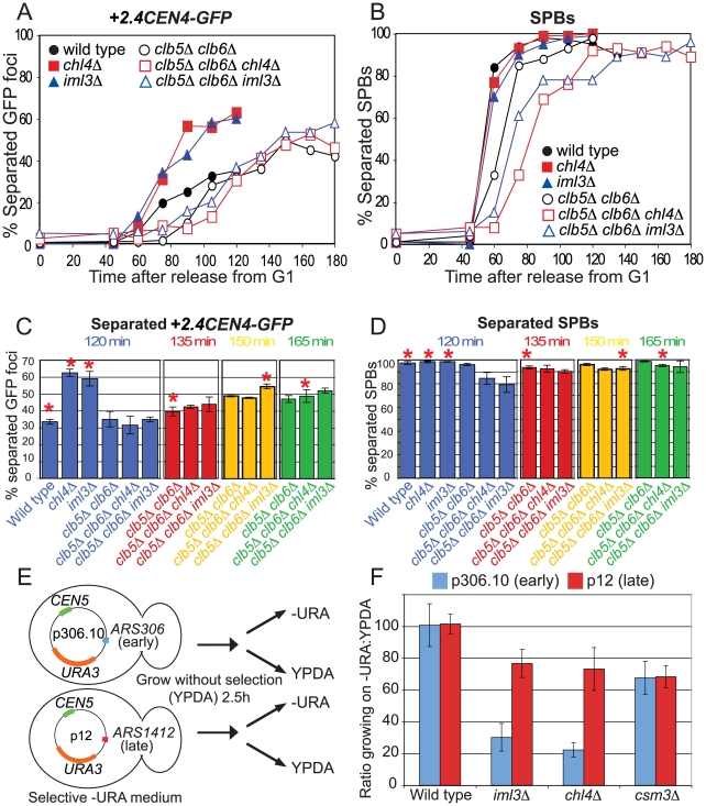
The cohesin complex holds sister chromatids together from the time of their duplication in S phase until their separation during mitosis. Although cohesin is found along the length of chromosomes, it is most abundant at the centromere and surrounding region, the pericentromere. We show here that the budding yeast Ctf19 kinetochore subcomplex and the replication fork-associated factor, Csm3, are both important mediators of pericentromeric cohesion, but they act through distinct mechanisms. We show that components of the Ctf19 complex direct the increased association of cohesin with the pericentromere. In contrast, Csm3 is dispensable for cohesin enrichment in the pericentromere but is essential in ensuring its functionality in holding sister centromeres together. Consistently, cells lacking Csm3 show additive cohesion defects in combination with mutants in the Ctf19 complex. Furthermore, delaying DNA replication rescues the cohesion defect observed in cells lacking Ctf19 complex components, but not Csm3. We propose that the Ctf19 complex ensures additional loading of cohesin at centromeres prior to passage of the replication fork, thereby ensuring its incorporation into functional linkages through a process requiring Csm3.


Conflict of interest statement

The authors have declared that no competing interests exist.

Figures

Figure 1
Figure 1. Analysis of Scc1 localization at the pericentromere and the effect of tension in Ctf19 complex mutants.
(A) Schematic diagram of the Ctf19 kinetochore subcomplex adapted from ,. (B) Schematic diagram showing locations of primers used for qPCR analysis of ChIP samples. The hatched area represents the core centromere, and the black, chromosome arms. The ∼50 kb pericentromere is split into white, representing the ∼20 kb region in which cohesin is displaced and sister chromatids separate under tension. The remainder of the pericentromere in which cohesin is retained under tension is shown in grey. (C) Analysis of Scc1-6HA association in cells arrested in metaphase of mitosis in the absence of microtubules. Strains AM1145 (SCC1-6HA), AM3441 (iml3Δ SCC1-6HA), AM3442 (chl4Δ SCC1-6HA), and AM1176 (no tag) were arrested in medium containing nocodazole and benomyl for 3 h to depolymerize microtubules and induce a metaphase arrest. (D) Analysis of Scc1-6HA association in cells arrested in metaphase of mitosis with sister kinetochores under tension. Strains AM1105 (SCC1-6HA), AM3948 (iml3Δ SCC1-6HA), AM3950 (chl4Δ SCC1-6HA), and AM2508 (no tag control) all carrying MET-CDC20 were arrested in G1 using alpha factor and released into medium containing methionine to deplete CDC20. Cells were harvested 2 hours after release from G1 and metaphase arrest confirmed. The mean values from three independent experiments are shown with error bars indicating standard deviation in (C) and (D).
Figure 2
Figure 2. Ctf19 complex mutants exhibit pericentromeric cohesion defects in mitosis.
(A) Sister centromeres (+2.4CEN4-GFP; 2.4 kb to the right of CEN4) are more frequently separated in a metaphase arrest in iml3Δand chl4Δ mutants and this depends on microtubules. Strains of wild type (AM914), iml3Δ (AM3522), and chl4Δ (AM3501), all carrying +2.4CEN4-GFP (2.4 kb to the right of CEN4) and MET-CDC20 were arrested in alpha factor and then released into medium containing methionine to deplete CDC20 and either DMSO (control) or nocodazole (to depolymerize microtubules). Samples were taken at the indicated times after release from G1 for anti-tubulin immunofluorescence (Figure S2) and to determine the number of GFP foci per nucleus. Percentages of cells with two GFP dots are shown for wild type (black squares), chl4Δ (blue circles), and iml3Δ (red triangles) treated with either DMSO (open symbols) or nocodazole (closed symbols). 200 cells were scored at each time point. (B) Cohesion defects were examined after releasing cells carrying +2.4CEN4-GFP, SPC42-tdTomato (to label spindle pole bodies) and MET-CDC20 from a G1 block, as a result of alpha factor treatment, into medium containing methionine to deplete CDC20. A snapshot of cells at the 120 min time point (metaphase arrest) is shown as an example together with a schematic diagram of chromosome IV with sister centromeres under tension (left). SPBs are shown in red and +2.4CEN4-GFP is shown in green. The black and white arrowheads indicate cohesed and separated +2.4CEN4-GFP foci, respectively. (C) Summary of frequency of +2.4CEN4-GFP separation at metaphase in Ctf19 complex mutants. Values are the mean of all experiments where percentages of separated sister centromeres were determined after scoring 200 cells, 120 min after release from G1, in a metaphase arrest. Error bars indicate standard error. Strains, and number of independent repeats (given in italics), were: wild type (AM4643; 14), chl4Δ (AM4644; 10), iml3Δ (AM4647; 7), ctf3Δ (AM4683; 3), mcm16Δ (AM6158; 2), mcm22Δ (AM6160; 2), ctf19Δ (AM5786; 3), mcm21Δ (AM5788; 3), chl4Δ mcm16Δ (AM6195; 1), chl4Δ mcm22Δ (AM6192, 1), and csm3Δ (AM4717; 6). (D,E) Sister centromeres and SPBs are further apart at metaphase in iml3Δ, chl4Δ, and ctf3Δ mutants. The distance between +2.4CEN4-GFP (D) and SPC42-tdTomato (E) foci was determined for all separated foci from the 60–120 minute time points. For wild type (AM4643), chl4Δ (AM4644), and iml3Δ (AM4647), the average of two independent experiments with error bars representing standard deviation is shown. For ctf3Δ (AM4683), measurements are from a single experiment. (D) The percentage of +2.4CEN4-GFP foci that were >0–1 µm (blue) or >1–4 µm (red) apart. n = 115/82 (wild type); 202/139 (chl4Δ), 184/167 (iml3Δ); 199 (ctf3Δ). (E) The percentage of SPC42-tdTomato foci that were >0–3 µm (blue) or >3–6 µm (red) apart is shown. n = 338/305 (wild type); 548/244 (chl4Δ), 331/272 (iml3Δ); 313 (ctf3Δ). The percentages of separated spindle pole bodies or +2.4CEN4-GFP foci are shown in Figure S2 for this experiment. (F–K) The cohesion defect in iml3Δ and chl4Δ mutants is not chromosome-specific but is restricted to centromere-proximal regions. Cells of the indicated genotypes carrying MET-CDC20, SPC42-tdTomato and with GFP labels at various loci were arrested in G1 with alpha factor and released into medium containing methionine and the percentages of separated GFP foci were scored at the indicated timepoints. tet operators are integrated at: (F) +4.5CEN6-GFP (4.5 kb to right of CEN6) in wild type (AM5329) and iml3Δ (AM5330); (G) +1.4CEN5-GFP (1.4 kb to right of CEN5) in wild type (AM5189), iml3Δ (AM5249), chl4Δ (AM5251), and ctf3Δ (AM5188); (H) -12.6CEN5-GFP (12.6 kb to left of CEN5) in wild type (AM5545), iml3Δ (AM5542), and chl4Δ (AM5560); (I) -17.8CEN5-GFP (17.8 kb to left of CEN5) in wild type (AM5533), iml3Δ (AM5537) and chl4Δ (AM5551); (J) URA3-GFP (38.4 kb to left of CEN5) in wild type (AM1081) and iml3Δ (AM3541) and chl4Δ (AM3519). (K) -30RTEL5 (∼30 kb from telomere on right arm of chromosome V) in wild type (AM2511) and iml3Δ (AM3887) and chl4Δ (AM3942). Note that the URA3-GFP and TEL5-GFP strains did not carry SPC42-tdTomato. Distances indicate the start of the tetO array from the centromere.
Figure 3
Figure 3. The Ctf19 complex promotes cohesion establishment in the pericentromere during meiosis.
Schematic diagram showing the segregation of chromosome V with either heterozygous (A) or homozygous (B) GFP labels in wild type cells. Homologs are shown in white and grey and the GFP dot is shown as a black circle. Sister chromatid cohesion is represented by black lines between sister chromatids. Arrows on kinetochores indicate the direction of attachment to the spindle. (C,D) Ctf19 complex mutations lead to errors in chromosome segregation during meiosis. Cells of the indicated genotypes in which either one copy [(C); heterozygous) or both copies (D); homozygous] of chromosome V were marked with GFP (tetR-GFP::LEU2 URA3::tetOx224) at URA3 (38.4 kb from the centromere) were induced to sporulate at 30°C. The percentage of tetranucleate cells with the patterns of GFP dot segregation shown was determined after examining 200 cells, 8 hours after transfer into sporulation medium. Strains used were AM107 (wild type), AM1904 (iml3Δ), AM1902 (chl4Δ), AM5104 (ctf3Δ), AM5105 (mcm16Δ), AM3684 (mcm22Δ), AM5107 (ctf19Δ), AM436 (mcm21Δ), AM4781 (nkp1Δ), and AM4988 (nkp2Δ) for (C) and AM1603 (wild type), AM1903 (iml3Δ), AM1905 (chl4Δ), AM3811 (iml3Δ chl4Δ), AM4059 (ctf3Δ), AM3799 (mcm16Δ), AM3661 (mcm22Δ), AM3798 (ctf19Δ), AM437 (mcm21Δ), and AM5809 (chl4Δ ctf3Δ) for (D). Values shown are the mean from two independent experiments, with error bars representing standard deviation, with the exception of mcm22Δ, nkp1Δ, nkp2Δ (C) and mcm22Δ and chl4Δ ctf3Δ (D), where results are shown from a single experiment. (E,F) Reduced levels of the meiotic cohesin, Rec8, at the pericentromere in Ctf19 complex mutants. (E) Schematic diagram of the primer sets used for qPCR on chromosome IV. (F) Analysis of Rec8-3HA association in cells arrested in metaphase I in meiosis. Strains AM3560 (no tag control), AM3375 (REC8-3HA), AM3422 (chl4Δ REC8-3HA) and AM3377 (iml3Δ REC8-3HA) carrying the pCLB2-3HA-CDC20 allele were harvested for ChIP 8 h after transfer into sporulation medium. The schematic diagram shows the configuration of the chromosomes. qPCR analysis of anti-HA ChIP samples was performed using primers shown in (E).
Figure 4
Figure 4. Csm3 promotes cohesion establishment at the pericentromere but is not required for cohesin enrichment in this region.
(A–E) csm3Δ mutants show cohesion defects in mitosis that are additive with iml3Δ. Strains carrying the indicated GFP labels and MET-CDC20 were released from G1 into a metaphase arrest by CDC20 depletion, and the number of GFP foci per nucleus was determined for 200 cells. Strains with +2.4CEN4-GFP, -12.6CEN5-GFP, and -17.8CEN5-GFP also carried SPC42-tdTomato. (A) Separation of +2.4CEN4-GFP foci in wild type (AM4643), csm3Δ (AM4717), and ctf3Δ (AM4683). (B) Separation of -12.6CEN5-GFP foci in wild type (AM5545), csm3Δ (AM5569), and chl4Δ (AM5560). (C) Separation of -17.8CEN5-GFP foci in wild type (AM5533), csm3Δ (AM5564), and chl4Δ (AM5551). (D) Separation of URA3-GFP foci in wild type (AM1081), chl4Δ (AM3519), and csm3Δ (AM5312). (E) Separation of URA3-GFP foci in wild type (AM1081), iml3Δ (AM3541), csm3Δ (AM5312), and csm3Δ iml3Δ (AM5796). (F) Summary of frequency of URA3-GFP foci separation at the 120 min timepoint. Values are the mean from 3 (wild type, iml3Δ, csm3Δ) or 2 (iml3Δ csm3Δ) experiments with error bars indicating standard error. For each experiment 200 cells were scored. (G,H) Csm3 is not required for cohesin enrichment in the pericentromere. Strains AM1145 (SCC1-6HA), AM4927 (ctf3Δ SCC1-6HA), AM3757 (csm3Δ SCC1-6HA), and AM1176 (no tag) were arrested in medium containing nocodazole and benomyl for 3 h to depolymerize microtubules and induce a metaphase arrest. Primers at locations depicted in (G) were used for analysis of Scc1-6HA association by qPCR after ChIP (H). (I,J) Deletion of RRM3 can partially rescue the cohesion defect of csm3Δ mutants. Separation of +2.4CEN4-GFP foci in wild type (AM4643), csm3Δ (AM4717), rrm3Δ (AM6068), and csm3Δ rrm3Δ (AM6066) in a representative experiment (I) and mean values at 120 mins from 3 independent experiments with error bars indicating standard error (J).
Figure 5
Figure 5. Chl4 directs cohesin association with the pericentromere throughout the cell cycle.
(A–E) Time course of cohesin loading in the cell cycle. Wild type (AM4226) and chl4Δ (AM4291) strains carrying MET-CDC20 and SCC1-6HA were arrested in G1 with alpha factor then released into medium containing methionine and nocodazole at 18°C. Samples were taken at the indicated timepoints and Scc1-6HA association was analyzed by ChIP and qPCR. (A) Schematic diagram of sites used for qPCR. Results for centromeric sites C1 (B), C2 (C), pericentromeric site P2 (D), and arm site A1 (E) are shown. Where error bars are present, the mean of 3 (C,E) or 2 (B,D) independent experiments are shown with error bars representing standard deviation. Time points without error bars where taken in only one of the experiments. (F–J) Chl4 is required for Scc1 to associate efficiently with the pericentromere during metaphase after the eradication of tension. (F) Schematic diagram showing the experimental set-up. (G,H) Strains AM1105 (SCC1-6HA), AM3950 (chl4Δ SCC1-6HA), and AM2508 (no tag control), carrying MET-CDC20 were arrested in G1 and then released into medium containing methionine for 90 min to induce a metaphase arrest in the presence of microtubules. Half the culture was harvested for Scc1-6HA ChIP analysis and nocodazole was added to the remainder to depolymerize microtubules. After 60 min the nocadazole-treated sample was harvested for ChIP analysis. (G). qPCR analysis after ChIP prior to nocodazole addition. (H) qPCR analysis after ChIP following microtubule depolymerization. (I,J) Chl4 contributes to the de novo loading of cohesin at the centromere during a metaphase arrest. Strains AM4084 (pGAL-SCC1-3HA) and AM5974 (chl4Δ pGAL-SCC1-3HA) were treated as described in (G,H) except that cells were arrested in metaphase by CDC20 depletion in raffinose, rather than glucose medium, and galactose was added to half of the culture, together with nocodazole, to induce SCC1-3HA expression. (I) qPCR analysis after ChIP prior to nocodazole and galactose addition. (J) qPCR analysis after ChIP following microtubule depolymerization and SCC1-3HA induction.
Figure 6
Figure 6. Delaying DNA replication by hydroxyurea treatment rescues the cohesion defect of iml3Δ and chl4Δ, but not csm3Δ, mutants.
The experimental setup is shown in (A). Cells were released from G1 into medium containing 8 mM methionine in the presence of HU. After 1.5 h, the HU was washed out and a metaphase arrest was induced either by depletion of CDC20 or by nocodazole treatment. (B–E) Analysis of +2.4CEN4-GFP foci separation after the HU-induced replication delay. Wild type (AM4643), chl4Δ (AM4644), iml3Δ (AM4647), ctf3Δ (AM4683), and csm3Δ (AM4717) strains were arrested in G1 with alpha factor, the cultures were split and released into 8 mM methionine either in the absence (B) or presence (C,D) of HU. Samples were taken at the indicated timepoints after release from G1 for scoring of +2.4CEN4-GFP (B,C) or SPC42-tdTomato (D) separation. FACS analysis was used to confirm a delay in bulk DNA replication in the HU-treated strains (Figure S9). (E) Mean GFP dot separation of 3 experiments at the 120 min time point after washing out HU. Error bars represent standard error. (F–H) Analysis of Scc1-6HA association with chromosome IV during metaphase with sister kinetochores not under tension, after an S phase delay. (F) Location of primer sets used for qPCR analysis. (G) Localization of Scc1 in the absence of microtubules. Strains used were AM1145 (SCC1-6HA), AM 3442 (chl4Δ SCC1-6HA), AM3757 (csm3Δ SCC1-6HA), and AM1176 (no tag). (H) Localization of Scc1 when sister kinetochores are under tension. qPCR analysis of Scc1-6HA ChIP from cells harvested 2 h after HU wash-out. Strains used all carried MET-CDC20 and were AM1105 (SCC1-6HA), AM3948 (iml3Δ SCC1-6HA), AM3950 (chl4Δ SCC1-6HA), and AM2508 (no tag control).
Figure 7
Figure 7. Replication timing affects cohesion establishment and plasmid maintenance in chl4Δ and iml3Δ mutants.
(A–D) The frequency of +2.4CEN4-GFP dot separation in clb5Δ clb6Δ cells is not greatly increased by deletion of IML3 or CHL4. Wild type (AM4643), chl4Δ (AM4644), iml3Δ (AM4647), clb5Δ clb6Δ (AM5351), clb5Δ clb6Δ chl4Δ (AM5426), and clb5Δ clb6Δ iml3Δ (AM5428) carrying MET-CDC20, +2.4CEN4-GFP, and SPC42-tdTomato were arrested in G1 with alpha factor and released into medium containing methionine. Percentages of separated GFP foci (A) and SPBs (B) are shown at the indicated time points for a representative experiment. After 120 min there were 5% (wild type), 3% (chl4Δ), and 1% (iml3Δ) anaphase cells, after which they escape from the metaphase arrest. Escape is delayed until 180 min in the clb5Δ clb6Δ cells, when anaphase cells account for 9% (clb5Δ clb6Δ), 3% (clb5Δ clb6Δ chl4Δ), and 9% (clb5Δ clb6Δ iml3Δ), respectively. Mean +2.4CEN4-GFP (C) and SPB (D) separation is shown from three (120, 135 min) or two (150, 165 min) experiments at the indicated timepoints with error bars representing standard error. (E,F) A plasmid with a late-replicating origin (p12) shows improved stability over a plasmid with an early-replicating origin (p306.10) in chl4Δ and iml3Δ cells. (E) Experimental outline is shown. (F). Ratio of colonies growing on selective medium that depends on the presence of the plasmid (-URA) compared to colonies growing on rich medium for wild type (AM1176), iml3Δ (AM3313), chl4Δ (AM3314), and csm3Δ (AM3194) strains transformed with the indicated plasmids.

References

    1. Nasmyth K. Disseminating the genome: joining, resolving, and separating sister chromatids during mitosis and meiosis. Annu Rev Genet. 2001;35:673–745. - PubMed
    1. Ciosk R, Shirayama M, Shevchenko A, Tanaka T, Toth A, et al. Cohesin's binding to chromosomes depends on a separate complex consisting of Scc2 and Scc4 proteins. Mol Cell. 2000;5:243–254. - PubMed
    1. Lengronne A, Katou Y, Mori S, Yokobayashi S, Kelly GP, et al. Cohesin relocation from sites of chromosomal loading to places of convergent transcription. Nature. 2004;430:573–578. - PMC - PubMed
    1. Uhlmann F, Nasmyth K. Cohesion between sister chromatids must be established during DNA replication. Curr Biol. 1998;8:1095–1101. - PubMed
    1. Katou Y, Kanoh Y, Bando M, Noguchi H, Tanaka H, et al. S-phase checkpoint proteins Tof1 and Mrc1 form a stable replication-pausing complex. Nature. 2003;424:1078–1083. - PubMed

Publication types

MeSH terms

Substances