. 2009 Sep;5(9):e1000569.
doi: 10.1371/journal.ppat.1000569. Epub 2009 Sep 4.

Plasmodium falciparum heterochromatin protein 1 marks genomic loci linked to phenotypic variation of exported virulence factors

Affiliations

Plasmodium falciparum heterochromatin protein 1 marks genomic loci linked to phenotypic variation of exported virulence factors

Christian Flueck et al. PLoS Pathog. 2009 Sep.

Abstract

Epigenetic processes are the main conductors of phenotypic variation in eukaryotes. The malaria parasite Plasmodium falciparum employs antigenic variation of the major surface antigen PfEMP1, encoded by 60 var genes, to evade acquired immune responses. Antigenic variation of PfEMP1 occurs through in situ switches in mono-allelic var gene transcription, which is PfSIR2-dependent and associated with the presence of repressive H3K9me3 marks at silenced loci. Here, we show that P. falciparum heterochromatin protein 1 (PfHP1) binds specifically to H3K9me3 but not to other repressive histone methyl marks. Based on nuclear fractionation and detailed immuno-localization assays, PfHP1 constitutes a major component of heterochromatin in perinuclear chromosome end clusters. High-resolution genome-wide chromatin immuno-precipitation demonstrates the striking association of PfHP1 with virulence gene arrays in subtelomeric and chromosome-internal islands and a high correlation with previously mapped H3K9me3 marks. These include not only var genes, but also the majority of P. falciparum lineage-specific gene families coding for exported proteins involved in host-parasite interactions. In addition, we identified a number of PfHP1-bound genes that were not enriched in H3K9me3, many of which code for proteins expressed during invasion or at different life cycle stages. Interestingly, PfHP1 is absent from centromeric regions, implying important differences in centromere biology between P. falciparum and its human host. Over-expression of PfHP1 results in an enhancement of variegated expression and highlights the presence of well-defined heterochromatic boundaries. In summary, we identify PfHP1 as a major effector of virulence gene silencing and phenotypic variation. Our results are instrumental for our understanding of this widely used survival strategy in unicellular pathogens.


Conflict of interest statement

The authors have declared that no competing interest exists

Figures
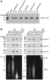
Figure 1
Figure 1. PfHP1 binds specifically to H3K9me3 and is associated with parasite heterochromatin.
(A) Peptide competition demonstrates the specific binding of PfHP1 to H3K9me3. Recombinant PfHP1-HIS bound to a biotinylated H3K9me3 peptide was immobilized on streptavidin agarose beads. PfHP1 was only eluted by competition with the H3K9me3 peptide (lane 3), whereas peptides H3K9me3S10p (lane 5), H3K9ac (lane 7) and H4K20me3 (lane 11) were unable to compete. PfHP1 had weak affinity for H3K27me3 (lane 9). After elution with peptides, remaining PfHP1-HIS was eluted with high salt (lanes 4, 6, 8, 10, 12). P, peptide elution; S, high salt elution; FT, Flow-through after coupling of PfHP1-HIS; wash, last wash prior to peptide elution. The Western blot was probed with anti-6×HIS antibodies. (B) Solubility of PfHP1, H3, H4, gDNA and RNA after treatment of parasite nulcei with nucleases. Isolated nuclei were extracted either with low-salt, MNAse, DNAse or RNAse, followed by serial treatment of the insoluble pellets with high-salt and SDS. Lanes 1–4: PfHP1 and H3/H4 were tightly associated with the salt-insoluble fraction. Partially degraded RNA and undigested gDNA were apparent in the high-salt and SDS-soluble fractions. Lanes 5–8: Digestion with MNAse solubilizes approx. 50% of PfHP1 and the entire pool of H3/H4. RNA was degraded by the single-stranded nuclease activity of MNAse. Mononucleosomes (MN) were completely extracted with 1 M KCl. Lanes 9–12: After digestion of nuclei with DNAseI, PfHP1 and H3/H4 extracted equally with high-salt and SDS. Genomic DNA was completely digested and partially degraded RNA extracted with high-salt and SDS. Lanes 13–16: Treatment with RNAse A did not affect the extractablity of PfHP1 and H3/H4. RNA was completely digested by RNAse A whereas intact gDNA remained associated with the insoluble fraction. Equal amounts were analysed for each protein and nucleic acid sample. RNA in ethidium bromide stained gels was identified by re-examination of the gel after incubation in RNAse A-containing buffer for 1 hr at RT. W2, second wash after extraction with 1 M KCl.
Figure 2
Figure 2. PfHP1 localizes to discrete regions at the nuclear periphery.
(A) PfHP1-GFP localization in unfixed 3D7/HP1-GFP parasites. (B) Localization of PfHP1-HA and (C) PfHP1-Ty in methanol-fixed parasites visualized by IFA. T, trophozoites. S, schizonts. (D) Localization of PfHP1-GFP by electron microsopy. Antibodies specific to GFP detect PfHP1-GFP at the nuclear periphery, reminiscent of the pattern seen by fluorescence microscopy (N, nucleus; C, cytoplasm). Schematic maps of the transfection constructs are shown above each panel.
Figure 3
Figure 3. PfHP1 associates with chromosome end clusters.
Combined IFA/FISH using 3D7/HP1-GFP ring stage parasites for simultaneous detection of chromosome end clusters and PfHP1-GFP. Fixed parasites were sequentially probed with anti-GFP antibodies (green) and the subtelomeric repeat sequence rep20 (TARE 6) (red). The distribution of GFP and rep20 signal numbers detected in 200 randomly selected nuclei and the frequency of GFP signals (659 in total) co-localising with, or directly adjacent to, rep20 signals are indicated.
Figure 4
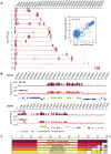
Figure 4. PfHP1 occupies virulence gene clusters throughout the genome and shows a high positive correlation with H3K9me3.
(A) Genome-wide PfHP1 occupancy as determined by high-density ChIP-on-chip. Schematic display (SignalMap) and localization of genomic regions bound by PfHP1-HA on all 14 chromosomes in 3D7/HP1-HA schizont stage parasites. PfHP1-HA occupancy for each gene was calculated as the average of log2 ratios of hybridization values for immunoprecipitated and input chromatin. Genome-wide H3K9me3 localization data obtained with the same NimbleGen platform are displayed on a negative scale. Chromosome numbers are indicated on the left, chromosomal positions on top. Green and black arrowheads identify PfHP1-bound loci that were either not enriched in H3K9me3 in two recent ChIP-on-chip studies, or for which no H3K9me3 data exist in the Lopez-Rubio study, respectively ,. The dot plot visualizes the high positive correlation between PfHP1 and H3K9me3 for all genes in the 3D7 genome (R2 = 0.72). (B) PfHP1-binding demarcates P. falciparum virulence gene clusters that are also enriched in H3K9me3. Regional zoom-in of ChIP-on-chip profiles of PfHP1 and H3K9me3 occupancy at a chromosome-central var gene cluster on chromosome 12 and the left end of chromosome 10. Chromosomal coordinates are according to PlasmoDB v5.5 annotation (www.plasmodb.org). PfHP1-bound gene types are color-coded below. (C) 387 of 425 PfHP1-bound genes are also enriched in H3K9me3 and are mostly members of lineage-specific exported gene families. Half of the 38 PfHP1-bound genes not enriched in H3K9me3 code for single copy genes. Clustering was performed according to information retrieved from PlasmoDB v5.5 (www.plasmodb.org) and the classification of exported proteins .
Figure 5
Figure 5. Validation of ChIP-on-chip results and mutually exclusive association of PfHP1/H3K9me3 and H3K9ac at P. falciparum virulence gene loci.
(A) Targeted ChIP-qPCR analysis demonstrates mutually exclusive presence of PfHP1/H3K9me3 and H3K9ac at selected loci. Recovery values of cross-linked PfHP1- and H3K9ac-associated chromatin from ten and twelve randomly selected loci tested negative or positive for PfHP1 in ChIP-on-chip, respectively, represent the mean from three independent experiments on schizont stage samples (twice 3D7/HP1-HA, once 3D7/HP1-GFP). Relative hybridisation intensities (RHI) from the ChIP-on-chip analysis are shown below each locus and indicate log2 ratios of recovered chromatin over input. (B) PfHP1 and H3K9ac profiles at the same loci in ring stage parasites are comparable to schizont stages. (C) H3K9me3-association in schizont stages was determined by ChIP-qPCR using non-cross-linked chromatin (native ChIP). Values derive from one of two independent experiments (3D7/HP1-HA and 3D7 wild-type) yielding similar results. qPCR primers (Table S4) targeted coding regions (open box) or non-coding promoter (solid line with arrow) or subtelomeric repeat (solid line) regions. Gene accession numbers are indicated below the bottom graph. Normal rabbit polyclonal IgG was used as negative control for ChIP.
Figure 6
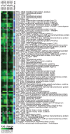
Figure 6. PfHP1 participates in silencing of variegated genes.
Significance Analysis for Microarrays (SAM) revealed 78 genes consistently down-regulated in two biological replicates (A and B) upon over-expression of PfHP1. The heat map compares the relative transcript abundances at four timepoints across the IDC (early ring stage (TP1), late ring stage (TP2), trophozoites (TP3), schizonts (TP4)) between the PfHP1-overexpressing and control lines. The right column highlights genes bound by PfHP1 in the ChIP-on-chip experiment (dark blue boxes), or genes that are members of PfHP1-bound gene families (light blue boxes). Gene accession numbers and annotations are according to PlasmoDB v5.5 (www.plasmodb.org).

References

    1. Snow RW, Guerra CA, Noor AM, Myint HY, Hay SI. The global distribution of clinical episodes of Plasmodium falciparum malaria. Nature. 2005;434:214–217. - PMC - PubMed
    1. Marti M, Baum J, Rug M, Tilley L, Cowman AF. Signal-mediated export of proteins from the malaria parasite to the host erythrocyte. J Cell Biol. 2005;171:587–592. - PMC - PubMed
    1. Sargeant TJ, Marti M, Caler E, Carlton JM, Simpson K, et al. Lineage-specific expansion of proteins exported to erythrocytes in malaria parasites. Genome Biol. 2006;7:R12. - PMC - PubMed
    1. MacPherson GG, Warrell MJ, White NJ, Looareesuwan S, Warrell DA. Human cerebral malaria. A quantitative ultrastructural analysis of parasitized erythrocyte sequestration. Am J Pathol. 1985;119:385–401. - PMC - PubMed
    1. Pongponratn E, Riganti M, Punpoowong B, Aikawa M. Microvascular sequestration of parasitized erythrocytes in human falciparum malaria: a pathological study. Am J Trop Med Hyg. 1991;44:168–175. - PubMed

Publication types

MeSH terms

Substances

Associated data