. 2009 Sep 4;4(9):e6893.
doi: 10.1371/journal.pone.0006893.

Ulcerative colitis induces changes on the expression of the endocannabinoid system in the human colonic tissue

Affiliations

Ulcerative colitis induces changes on the expression of the endocannabinoid system in the human colonic tissue

Lucia Marquéz et al. PLoS One. .

Abstract

Background: Recent studies suggest potential roles of the endocannabinoid system in gastrointestinal inflammation. Although cannabinoid CB(2) receptor expression is increased in inflammatory disorders, the presence and function of the remaining proteins of the endocannabinoid system in the colonic tissue is not well characterized.

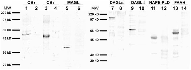
Methodology: Cannabinoid CB(1) and CB(2) receptors, the enzymes for endocannabinoid biosynthesis DAGLalpha, DAGLbeta and NAPE-PLD, and the endocannabinoid-degradating enzymes FAAH and MAGL were analysed in both acute untreated active ulcerative pancolitis and treated quiescent patients in comparison with healthy human colonic tissue by immunocytochemistry. Analyses were carried out according to clinical criteria, taking into account the severity at onset and treatment received.

Principal findings: Western blot and immunocytochemistry indicated that the endocannabinoid system is present in the colonic tissue, but it shows a differential distribution in epithelium, lamina propria, smooth muscle and enteric plexi. Quantification of epithelial immunoreactivity showed an increase of CB(2) receptor, DAGLalpha and MAGL expression, mainly in mild and moderate pancolitis patients. In contrast, NAPE-PLD expression decreased in moderate and severe pancolitis patients. During quiescent pancolitis, CB(1), CB(2) and DAGLalpha expression dropped, while NAPE-PLD expression rose, mainly in patients treated with 5-ASA or 5-ASA+corticosteroids. The number of immune cells containing MAGL and FAAH in the lamina propria increased in acute pancolitis patients, but dropped after treatment.

Conclusions: Endocannabinoids signaling pathway, through CB(2) receptor, may reduce colitis-associated inflammation suggesting a potential drugable target for the treatment of inflammatory bowel diseases.


Conflict of interest statement

Competing Interests: The authors have declared that no competing interests exist.

Figures

Figure 1
Figure 1. Western blots of membrane extracts from human colonic tissue.
They showed prominent immunoreactive bands of the expected size for the ECS proteins. See text. Positions of molecular markers (MW) are indicated at the left.
Figure 2
Figure 2. Immunohistochemistry for CB1 and CB2 receptors, FAAH and MAGL in human colonic tissue.
Morphology of normal human colon, stained with H&E (A–C). General views of transmural sections through the colon (A, D, G, J, M). High-magnification photomicrographs of the colonic epithelium and lamina propria (B, E, H, K, N), smooth muscle and myenteric plexus (C, F, I, L, O). Abbreviations: C, crypt; CSM, circular smooth muscle; LP, lamina propria; LSM, longitudinal smooth muscle; M, mucosa; MM, muscularis mucosae, MP, myenteric plexus; SM, submucosa.
Figure 3
Figure 3. Immunohistochemistry for NAPE-PLD, DAGLα and DAGLβ in human colonic tissue.
General views of transmural sections through the colon (A, D, G). High-magnification photomicrographs of the colonic epithelium and lamina propria (B, E, H), smooth muscle and myenteric plexus (C, F, I).
Figure 4
Figure 4. Immunohistochemistry in human healthy (control), acute UC and quiescent UC colonic tissue.
Representative microphotographs of CB1 receptor (A–C), CB2 receptor (D–F), DAGLα (G–I), DAGLβ (J–L), MAGL (M–O), NAPE-PLD (P–R), FAAH (S–V) were shown.
Figure 5
Figure 5. Quantification of ECS component immunoreactivity in the colonic epithelium.
A: Untreated acute UC at disease onset showed increases in CB2, DAGLα and MAGL immunoreactivity, and decreases in NAPE-PLD immunostaining. After achieving remission (quiescence), CB1 and CB2 receptor immunoreactivity dropped, MAGL immunostaining maintained the same levels than acute group and NAPE-PLD immunoreactivity reverted to control levels. B: CB2/CB1 ratio increased in both groups. However, CB2 immunoreactivity increased in acute patients, while in quiescent patients there was a decrease of CB1 receptor and a reverted restoration of CB2 level. NAPE-PLD/FAAH ratio dropped in acute group, but rose to control levels in quiescent one. Histograms represent the mean±SEM. U Mann Witney and Wilcoxon tests: *P<0.05, **P<0.01 and ***P<0.001 versus control group; #P<0.05 and ##P<0.01 versus acute group. N = 22, 24 and 24 for control, acute and quiescent groups respectively.
Figure 6
Figure 6. Percentage of immunoreactive immune cells for ECS components in the lamina propria.
Untreated acute UC at disease onset is associated with high number of FAAH+ and MAGL+ immune cells that was significantly diminished after treatment only in FAAH immunoreactivity. Histograms represent the mean±SEM. U Mann Witney and Wilcoxon tests: ***P<0.001 versus control group; ###P<0.001 versus acute group. N = 22, 24 and 24 for control, acute and quiescent groups respectively.
Figure 7
Figure 7. Quantification of epithelial ECS immunoreactivity depending on the UC severity score.
Main changes were observed mainly in mild and moderate acute UC. Histograms represent the mean±SEM. U Mann Witney and Wilcoxon tests: *P<0.05, **P<0.01 and ***P<0.001 versus control group; #P<0.05 versus acute group. N = 6, 13 and 5 for mild, moderate and severe groups respectively.
Figure 8
Figure 8. Quantification of epithelial ECS immunoreactivity depending on the treatment received.
As a relevant finding, treatment is associated with changes in the expression of cannabinoid receptors and EC-production and -degradation enzymes, suggesting that these proteins can be considered as biomarkers of active disease/response to treatment. Histograms represent the mean±SEM. U Mann Witney and Wilcoxon tests: *P<0.05 and **P<0.01 versus control group; #P<0.05 versus acute group. N = 3, 15 and 6 for 5-ASA, 5-ASA+corticosteroids and 5-ASA+corticosteroids+immunomodulators respectively.

References

    1. Massa F, Storr M, Lutz B. The endocannabinoid system in the physiology and pathophysiology of the gastrointestinal tract. J Mol Med. 2005;83:944–954. - PubMed
    1. Pinto L, Capasso R, Di Carlo G, Izzo AA. Endocannabinoids and the gut. Prostag Leukotr Ess. 2002;66:333–341. - PubMed
    1. Pertwee RG. Cannabinoids and the gastrointestinal tract. Gut. 2001;48:859–867. - PMC - PubMed
    1. Izzo AA, Mascolo N, Capasso F. The gastrointestinal pharmacology of cannabinoids. Curr Opin Pharmacol. 2001;1:597–603. - PubMed
    1. Fowler CJ, Holt S, Nilsson O, Jonsson KO, Tiger G, et al. The endocannabinoid signaling system: pharmacological and therapeutic aspects. Pharmacol Biochem Behav. 2005;81:248–262. - PubMed

Publication types

Substances