. 2009 Sep 4;4(9):e6908.
doi: 10.1371/journal.pone.0006908.

Homeostatic plasticity of striatal neurons intrinsic excitability following dopamine depletion

Affiliations

Homeostatic plasticity of striatal neurons intrinsic excitability following dopamine depletion

Karima Azdad et al. PLoS One. .

Abstract

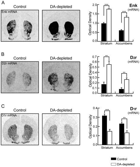
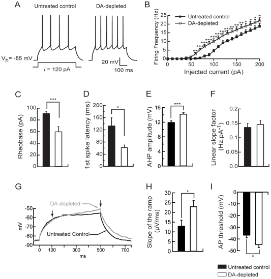
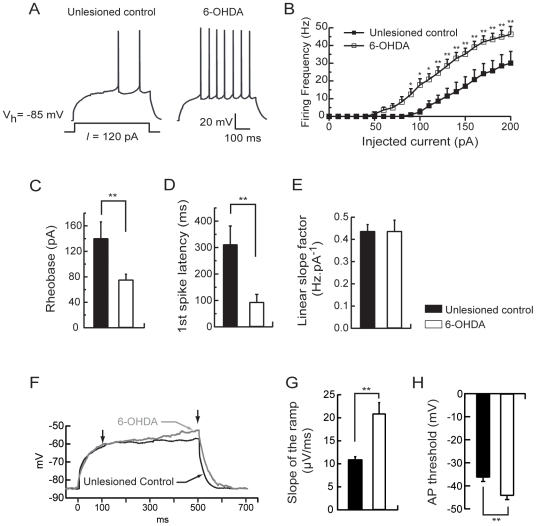
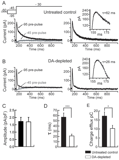
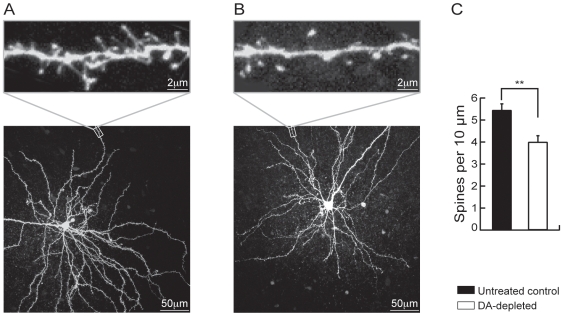
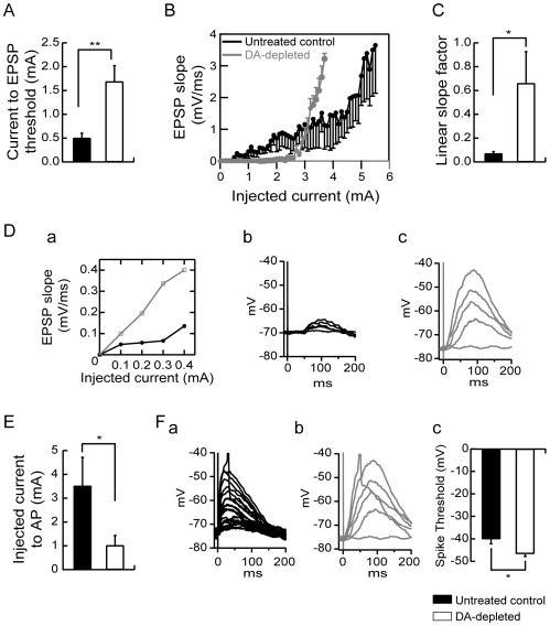
The striatum is the major input structure of basal ganglia and is involved in adaptive control of behaviour through the selection of relevant informations. Dopaminergic neurons that innervate striatum die in Parkinson disease, leading to inefficient adaptive behaviour. Neuronal activity of striatal medium spiny neurons (MSN) is modulated by dopamine receptors. Although dopamine signalling had received substantial attention, consequences of dopamine depletion on MSN intrinsic excitability remain unclear. Here we show, by performing perforated patch clamp recordings on brain slices, that dopamine depletion leads to an increase in MSN intrinsic excitability through the decrease of an inactivating A-type potassium current, I(A). Despite the large decrease in their excitatory synaptic inputs determined by the decreased dendritic spines density and the increase in minimal current to evoke the first EPSP, this increase in intrinsic excitability resulted in an enhanced responsiveness to their remaining synapses, allowing them to fire similarly or more efficiently following input stimulation than in control condition. Therefore, this increase in intrinsic excitability through the regulation of I(A) represents a form of homeostatic plasticity allowing neurons to compensate for perturbations in synaptic transmission and to promote stability in firing. The present observations show that this homeostatic ability to maintain firing rates within functional range also occurs in pathological conditions, allowing stabilizing neural computation within affected neuronal networks.


Conflict of interest statement

Competing Interests: The authors have declared that no competing interests exist.

Figures

Figure 1
Figure 1. Dopamine depletion changes expression of specific striatal genes.
Left panels, Autoradiograms generated by in situ hybridization using α-35S-labeled-labeled oligoprobes to identify enkephalin (A), dopamine D2 receptor (B) and dopamine D1 receptor (C) mRNA expression from untreated control and dopamine-depleted animals. Right panels, Levels of expression of enkephalin mRNA (A), D2 receptor mRNA (B) and D1 receptor mRNA (C) in striatum and nucleus accumbens in both conditions. (Untreated control rats n = 5, dopamine (DA)-depleted rats n = 6; data represent mean ± SEM expressed in optical density, arbitrary unit; **p<0.01, ***p<0.001).
Figure 2
Figure 2. Dopamine depletion increases intrinsic excitability of medium spiny neurons.
(A) Representatives traces showing that evoked action potentials (120 pA current pulse) were increased in dopamine-depleted MSN. (B) Summary plot of discharge frequency as a function of injected current illustrating the effect of dopamine depletion on the firing frequency. Dopamine depletion significantly shifted the curve to the left. (C) Histogram of the mean rheobase values showing the decrease of the average minimal depolarizing current amplitude required to elicit spike discharge (rheobase) in dopamine-depleted MSN compared to untreated controls. (D) Summary histogram of the mean first spike latency at the 120 pA current pulse illustrating the decrease of the first spike latency induced by dopamine depletion. (E) Histogram illustrating the effect of dopamine depletion on AHP amplitude at the 120 pA current pulse. (F) Histogram values of mean linear slope of the current-frequency plot in both conditions. (G) Representatives traces from untreated control and dopamine-depleted conditions, show the slowly developing ramp potential preceding firing. The slope of the ramp membrane potential was estimated between 100 and 500 ms after the induction of the 500 ms-depolarising step (arrows). (H) The increase of the slope of the ramp in dopamine-depleted condition is illustrated in the summary histogram. (I) Histogram values of the action potential threshold in dopamine-depleted MSN compared to untreated controls. (Untreated control medium spiny neurons n = 8, dopamine (DA)-depleted medium spiny neurons n = 8; data represent mean ± SEM; *p<0.05, **p<0.01, ***p<0.001).
Figure 3
Figure 3. 6-OHDA lesion-induced dopamine depletion increases intrinsic excitability of medium spiny neurons.
(A) Representatives traces showing that evoked action potentials (120 pA current pulse) were increased in MSN from 6-OHDA lesioned animals. (B) Summary plot of discharge frequency as a function of injected current illustrating the effect of 6-OHDA lesion on the firing frequency. 6-OHDA lesion-induced dopamine depletion significantly shifted the curve to the left. (C) Histogram of the mean rheobase values showing the decrease of the average minimal depolarizing current amplitude required to elicit spike discharge (rheobase) in MSN from 6-OHDA lesioned animals compared to unlesioned controls. (D) Summary histogram of the mean first spike latency at the 120 pA current pulse illustrating the decrease of the first spike latency induced by 6-OHDA lesion. (E) Histogram values of mean linear slope of the current-frequency plot in both conditions. (F) Representatives traces from MSN in unlesioned control and 6-OHDA lesioned conditions, show the slowly developing ramp potential preceding firing. The slope of the ramp membrane potential was estimated between 100 and 500 ms after the induction of the 500 ms-depolarising step (arrows). (G) The increase of the slope of the ramp in 6-OHDA lesioned condition is illustrated in the summary histogram. (H) Histogram values of the action potential threshold in MSN from 6-OHDA lesioned animals compared to unlesioned controls. (medium spiny neurons from unlesioned control animals n = 7, medium spiny neurons from 6-OHDA lesioned animals n = 10; data represent mean ± SEM; *p<0.05, **p<0.01, ***p<0.001).
Figure 4
Figure 4. Dopamine depletion alters the A-type potassium current in MSN.
(A, B) Left column, Representative current traces obtained in each studied condition (control and dopamine depletion) and evoked by the voltage-clamp protocol shown on the top. Right column, A-type currents obtained from subtraction of currents traces of the left column (see text for details). Insets on the right column, Semilogarithmic plots of A-type current traces show that the current inactivation could be fit by a monoexponential function. Summary histograms of the mean current amplitude density (C), the inactivation time constant (τ) (D) and the total charge efflux (E) in untreated control and dopamine depleted MSN. These histograms illustrate the significant decreases of the inactivation time constant (τ) and total charge efflux induced by dopamine depletion. (Untreated control medium spiny neurons n = 8, dopamine (DA)-depleted medium spiny neurons n = 8; data represent mean ± SEM; *p<0.05, ***p<0.001).
Figure 5
Figure 5. Dopamine depletion reduces spines density in MSN.
Representative MSN in 280-µm-thick cortico-striatal slice from an untreated control rat (A) and a dopamine-depleted rat (B). Neurons were loaded with biocytin through the patch pipette and imaged by two-photon confocal microscopy. Maximum projection images of the soma and dendritic field (bottom panels A and B) and high-magnification projections of distal dendrite segments are shown (top panels A and B). (C) Histogram illustrating the effect of dopamine depletion on spine density. (Untreated control MSN n = 13, dopamine (DA)-depleted medium spiny neurons n = 7; data represent mean ± SEM; **p<0.01).
Figure 6
Figure 6. Dopamine depletion modulates excitatory synaptic transmission.
(A) Summary histogram of the mean minimal stimulation threshold current required to evoke an EPSP showing a significant increase induced by dopamine depletion. (B) Summary plot of EPSP slope as a function of increasing intensities of stimulation from untreated control and dopamine-depleted MSN. (C) Histogram of the mean slope factor ((mV/ms).mA−1) of the linear part of stimulation-EPSP slope plots in both groups. Dopamine depletion induced an increase in the strength of the excitatory fiber-MSN synapse. (D) Representative stimulation-EPSP slope plots (a) and traces of stimulation-evoked EPSPs recorded in current clamp from untreated control (black in (a) and (b)) and dopamine-depleted (gray in (a) and (c)) MSN. (E) Summary histogram illustrating the intensity of stimulation current needed to evoke an action potential above threshold current to evoke an EPSP. The intensity of current to action potential was significantly decreased in neurons from dopamine-depleted animals which is shown in representative traces from untreated control (Fa) and dopamine-depleted (Fb) MSN. The spike threshold was decreased in dopamine-depleted MSN compared to untreated controls as shown in the representative traces (Fa, Fb) and the values histogram (Fc). (Untreated control medium spiny neurons n = 7, dopamine (DA)-depleted medium spiny neurons n = 7; data represent mean ± SEM; *p<0.05, **p<0.01).

References

    1. Graybiel AM, Aosaki T, Flaherty AW, Kimura M. The basal ganglia and adaptive motor control. Science. 1994;265:1826–1831. - PubMed
    1. Nicola SM, Surmeier J, Malenka RC. Dopaminergic modulation of neuronal excitability in the striatum and nucleus accumbens. Annu Rev Neurosci. 2000;23:185–215. - PubMed
    1. Shen W, Flajolet M, Greengard P, Surmeier DJ. Dichotomous dopaminergic control of striatal synaptic plasticity. Science. 2008;321:848–851. - PMC - PubMed
    1. Surmeier DJ, Ding J, Day M, Wang Z, Shen W. D1 and D2 dopamine-receptor modulation of striatal glutamatergic signaling in striatal medium spiny neurons. Trends Neurosci. 2007;30:228–235. - PubMed
    1. Calabresi P, Centonze D, Bernardi G. Electrophysiology of dopamine in normal and denervated striatal neurons. Trends Neurosci. 2000;23:S57–63. - PubMed

Publication types