. 2009 Dec;8(6):676-89.
doi: 10.1111/j.1474-9726.2009.00517.x. Epub 2009 Sep 2.

Relative roles of TGF-beta1 and Wnt in the systemic regulation and aging of satellite cell responses

Affiliations
Free PMC article

Relative roles of TGF-beta1 and Wnt in the systemic regulation and aging of satellite cell responses

Morgan E Carlson et al. Aging Cell. 2009 Dec.
Free PMC article

Abstract

Muscle stem (satellite) cells are relatively resistant to cell-autonomous aging. Instead, their endogenous signaling profile and regenerative capacity is strongly influenced by the aged P-Smad3, differentiated niche, and by the aged circulation. With respect to muscle fibers, we previously established that a shift from active Notch to excessive transforming growth factor-beta (TGF-beta) induces CDK inhibitors in satellite cells, thereby interfering with productive myogenic responses. In contrast, the systemic inhibitor of muscle repair, elevated in old sera, was suggested to be Wnt. Here, we examined the age-dependent myogenic activity of sera TGF-beta1, and its potential cross-talk with systemic Wnt. We found that sera TGF-beta1 becomes elevated within aged humans and mice, while systemic Wnt remained undetectable in these species. Wnt also failed to inhibit satellite cell myogenicity, while TGF-beta1 suppressed regenerative potential in a biphasic fashion. Intriguingly, young levels of TGF-beta1 were inhibitory and young sera suppressed myogenesis if TGF-beta1 was activated. Our data suggest that platelet-derived sera TGF-beta1 levels, or endocrine TGF-beta1 levels, do not explain the age-dependent inhibition of muscle regeneration by this cytokine. In vivo, TGF-beta neutralizing antibody, or a soluble decoy, failed to reduce systemic TGF-beta1 and rescue myogenesis in old mice. However, muscle regeneration was improved by the systemic delivery of a TGF-beta receptor kinase inhibitor, which attenuated TGF-beta signaling in skeletal muscle. Summarily, these findings argue against the endocrine path of a TGF-beta1-dependent block on muscle regeneration, identify physiological modalities of age-imposed changes in TGF-beta1, and introduce new therapeutic strategies for the broad restoration of aged organ repair.


Figures

Fig. 3
Fig. 3
Old serum inhibits satellite cell responses by a Wnt-independent mechanism. Bioactive human (A) and mouse (B) Wnt is undetectable in human or mouse sera, and is not elevated with age. Human serum was collected from three young (∼20–25 years) and three old (∼65–75 years) individuals. Mouse serum was collected from four young and four aged mice. Levels of biologically active Wnt were analyzed, using a Wnt-reporter expressing cell line. As compared to recombinant Wnt3A, no detectable Wnt activity was found in either young or old serum. Raw fluorescence values for young sera were marginally higher compared to old sera, but both were not significantly different from the negative control (no Wnt3a samples). Inset panel shows 0.1–10 ng mL−1 Wnt3a range in greater detail. Myofiber-associated myogenic progenitor cells were isolated 3 days postinjury and cultured overnight in Opti-MEM containing either (C) 10% young serum (YS); YS+Wnt3A; YS+Wnt3A+TGF-β; or (E) 10% old (OS), OS+FRP3, OS+FRP3 + anti-TGF-β. BrdU was added for the last 2 h to measure proliferation. Cells were then fixed and immunostained for desmin (green) and BrdU (red), with Hoechst (blue) marking all nuclei. TGF-β addition reduced myoblast and myotube production in young serum, TGF-β neutralization resulted in a much improved myogenic proliferation in old serum, and some cells even formed de novo myotubes. In contrast, exogenous Wnt3A did not decrease myogenic responses in young serum and FRP3 did not rescue myogenic responses in old serum. No synergy in regulation of myogenesis was detected between Wnt and TGF-β. (D) Quantification of C. Cells were scored in multiple random fields from the above assays and the results displayed as the mean percent of BrdU+, desmin+/total cells, ±SD. *P < 0.05 between young untreated or +Wnt3a vs. +TGF-β or +TGF-β/Wnt3a; **P < 0.05 for old fibers, as described above for young. n = 3 for each set. (F) Quantification of E. Cells were scored and displayed as in E. *P < 0.05 between young fibers + OS untreated or +FRP3 vs. +anti-TGF-β or +anti-TGF-β/FRP3; **P < 0.05 for old fibers as described for young fibers; n = 3 for each set.
Fig. 1
Fig. 1
Old sera inhibits satellite cell responses by transforming growth factor (TGF)-β-dependent mechanism. Young (A) and old (B) myofiber-associated myogenic progenitor cells were isolated 3 days postinjury and cultured overnight in Opti-MEM containing either 10% young serum (YS), 10% old (OS), TGF-β1 antibody depleted serum alone, or with fixed amounts of recombinant TGF-β1 in the culture system. Cells were cultured with their specific sera for 24 h, and transferred to differentiation medium for additional 48 h (Fig S2). BrdU was added for the last 2 h to measure proliferation. Cells were then fixed and immunostained for desmin (green) and BrdU (red), with Hoechst (blue) marking all nuclei (as shown in C), and scored in multiple random fields from the above assays. Results are displayed as the mean percent of Desmin+/BrdU+/total cells, ±SD. *P < 0.05 for isochronic Y+YS/O+OS 0 ng mL−1, compared to 0.5 ng mL−1, and 5.0 ng mL−1 compared to 0.5 ng mL−1; n = 3.
Fig. 2
Fig. 2
Transforming growth factor (TGF)-β1 levels become elevated in old sera. (A) Mouse sera TGF-β1 becomes elevated with age. Mean serum levels, ±SEM, of TGF-β1 in young (Ym) or old (Om, n = 12 for each) mice as determined by sandwich ELISA; P < 0.0001. (B) Human TGF-β1 serum levels become elevated with age. TGF-β1 levels in the serum of aged humans (AG: 65–90 years old) vs. young (YG: 20–35 year old) were determined by ELISA. Error bars (SEM) represent the mean of 53 different subjects in each group; P < 0.0001. Similar age-related elevations in TGF-β1 were detected by bioactivity assay (Fig. S3). Dot plots represent separate animals (A) and separate individuals (D) of indicated ages. (C) Young and old myofiber explants were isolated at 3 days postinjury, and cultured overnight in Opti-MEM containing either young or old serum alone (control), or serum that had undergone activation of total TGF-β alone, in combination with neutralizing antibody to TGF-β1, or mixed with control serum. BrdU was added for the last 2 h to measure proliferation. Cells were fixed and immunostained for desmin (green) and BrdU (red), with Hoechst (blue) marking all nuclei (as shown in C, quantified in D). Cells were scored in multiple random fields and results are displayed as the mean percent of Desmin+/BrdU+ cells, ±SD. *P < 0.05 for activated compared to control, activated + antibody compared to activated, and control compared to activate + control. n = 3.
Fig. 4
Fig. 4
Down-modulation of transforming growth factor (TGF)-β signaling intensity by dominant negative TGF-β RII attenuates old sera-imposed satellite cell regenerative potential inhibition. (A) Young and old mice were injured with CTX, as described; bulk myofibers with activated satellite cells were then explanted and transiently transduced with a TGF-β RII DN-expressing lentivirus, or control virus, in vitro. Following transduction, myofiber-derived cells were cultured for 48 h in OPTI-MEM + 5% young (YS) or old (OS) mouse serum. Cells were fixed and analyzed by immunofluorescence for the expression of desmin (green), and levels of BrdU incorporation (red). Hoechst (blue) labels all nuclei. The successful down-modulation of TGF-β signaling by DN RII virus provided restoration of satellite cell regenerative potential in cells exposed to old serum, as evidenced by large number of de-novo proliferating desmin+ myoblasts that form multinucleated myotubes. Bar = 50 um. (B) Quantification of regenerative potential shown in A, The percent of desmin+/BrdU+ cells is shown as means with standard deviations. (*) indicates P < 0.05, compared to treatment with young serum and control virus (Y serum + cntl.) (**) indicates P < 0.05, as compared to treatment with old serum and control virus (O serum + cntl.). (C) Levels of nuclear P-Smad3 were diminished in old satellite cells transduced with DN RII-expressing lentivirus, in vitro. Old satellite cells, exposed to old serum (OS) plus control virus, or old serum plus DN RII-expressing virus (as indicated), were immunostained for P-Smad3 (green). Hoechst (blue) labels nuclei. As expected, levels of satellite cell nuclear P-Smad3 were diminished in cells transduced with DN RII-expressing virus, as compared to control (white arrow). Bar = 25 μm.
Fig. 5
Fig. 5
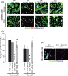
Systemic pharmacological intervention lowers sera transforming growth factor (TGF)-β levels and improves regeneration in old animals. Old or young mice were injected (subcutaneous) with a small molecule inhibitor of the TGF-β RI kinase (Ki), for 2 weeks. Five days before the end of treatment, muscle was injured as described. At the end of treatment, animals were sacrificed and muscle was collected. BrdU was injected (intraperitoneal) at 3 days postinjury to label proliferating, fusion-competent myoblasts. (A) Cryosections (10 μm) were performed in young and old muscle (receiving vehicle alone), old + Ki and old + RII (extracellular portion of TGF-β receptor II). Sections were analyzed by hematoxylin/eosin (H&E) staining and immunostaining for both embryonic myosin heavy chain (eMyHC, shown in green) and BrdU incorporation (shown in red). Hoechst stains nuclei (blue). As shown, the regenerative outcome of old and old + RII was worse than young (judged by scar tissue formation, de novo myofiber size/scarcity and reduction of BrdU+ nuclei within eMyHC fibers). Scale bar = 100 μm. In contrast, muscle repair was improved by treatment with TGF-β RI kinase (Ki) small molecule inhibitor (similar to young with respect to large/dense eMyHC+ myofibers with centrally located BrdU+ nuclei). (B) Regeneration was quantified from muscle sections, and is presented as mean percent of newly regenerated myofibers per square millimeter of injury site. Error bars indicate SD, n = 3, *P = 0.01 between young or old +Ki, and old.
Fig. 6
Fig. 6
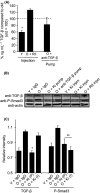
Systemic pharmacological intervention lowers sera transforming growth factor (TGF)-β levels and improves regeneration in old animals. (A) Myofibers were isolated from young, old and old mice with indicated treatments for TGF-β systemic down-modulation. Cell lysates were then analyzed by Western blot for levels of secretory TGF-β and P-Smad3 signaling strength. Actin immunodetection was used as a loading control. As expected, higher levels of TGF-β and P-Smad3 were detected in old samples, as compared to young. O + Ki pump showed reduction in both TGF-β and P-Smad3, whereas O + Ki injection only reduced P-Smad3 signaling strength. (B) Results of multiple Western blot assays were quantified (normalization of TGF-β and P-Smad3 pixel density by actin-specific pixel density) and are depicted by relative pixel intensity, as shown; (*) indicates P < 0.01, O Ki compared to old and young. Error bars indicate SD. n = 3–4. (C) TGF-β levels in serum for each animal in young, old or old + experimental treatments were determined by ELISA. Shown are mean values with standard deviations (n = 3–12, P ≤ 0.05 between O + Ki pump and O + anti-TGFβ pump, and O + Ki/O + RII injection.

References

    1. Abe M, Harpel JG, Metz CN, Nunes I, Loskutoff DJ, Rifkin DB. An assay for transforming growth factor-beta using cells transfected with a plasminogen activator inhibitor-1 promoter-luciferase construct. Anal. Biochem. 1994;216:276–284. - PubMed
    1. Andrews ZB, Zhao H, Frugier T, Meguro R, Grattan DR, Koishi K, McLennan IS. Transforming growth factor beta2 haploinsufficient mice develop age-related nigrostriatal dopamine deficits. Neurobiol. Dis. 2006;21:568–575. - PubMed
    1. Annes JP, Munger JS, Rifkin DB. Making sense of latent TGFbeta activation. J. Cell Sci. 2003;116:217–224. - PubMed
    1. Assoian RK, Komoriya A, Meyers CA, Miller DM, Sporn MB. Transforming growth factor-beta in human platelets. Identification of a major storage site, purification, and characterization. J. Biol. Chem. 1983;258:7155–7160. - PubMed
    1. Bonyadi M, Rusholme SA, Cousins FM, Su HC, Biron CA, Farrall M, Akhurst RJ. Mapping of a major genetic modifier of embryonic lethality in TGF beta 1 knockout mice. Nat. Genet. 1997;15:207–211. - PubMed

Substances