. 2009 Sep;158(2):610-20.
doi: 10.1111/j.1476-5381.2009.00341.x.

Modulation of cellular redox state underlies antagonism between oxaliplatin and cetuximab in human colorectal cancer cell lines

Affiliations

Modulation of cellular redox state underlies antagonism between oxaliplatin and cetuximab in human colorectal cancer cell lines

Laetitia Dahan et al. Br J Pharmacol. 2009 Sep.

Abstract

Background and purpose: Oxaliplatin is the first platinum-based compound effective in the treatment of colorectal cancer. Oxaliplatin combined with cetuximab for metastatic colorectal cancer is under evaluation. The preliminary results seem controversial, particularly for the use of cetuximab in K-Ras mutated patients. K-Ras mutation is known to affect redox homeostasis. Here we evaluated how the efficacy of oxaliplatin alone or combined with cetuximab varied according to the Ras mutation and redox status in a panel of colorectal tumour cell lines.

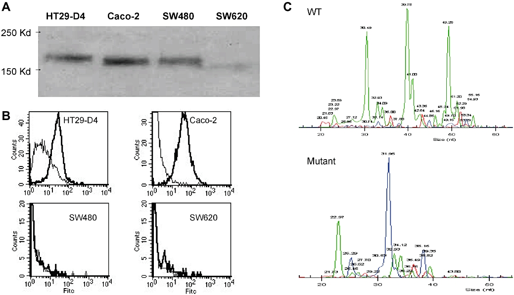
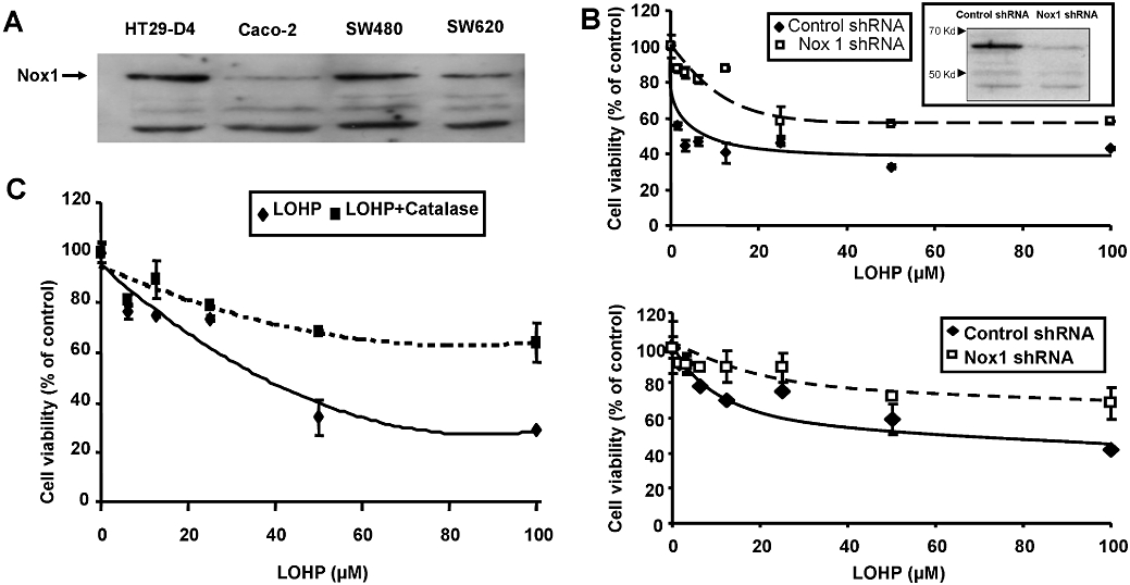
Experimental approach: Viability was evaluated by methylthiazoletetrazolium assay, reactive oxygen species production by DCFDA and lucigenin on HT29-D4, Caco-2, SW480 and SW620 cell lines.

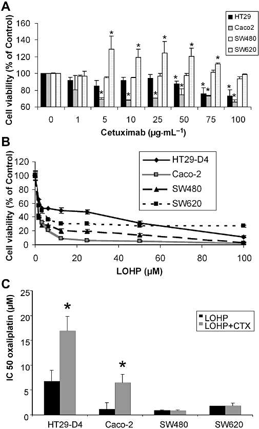
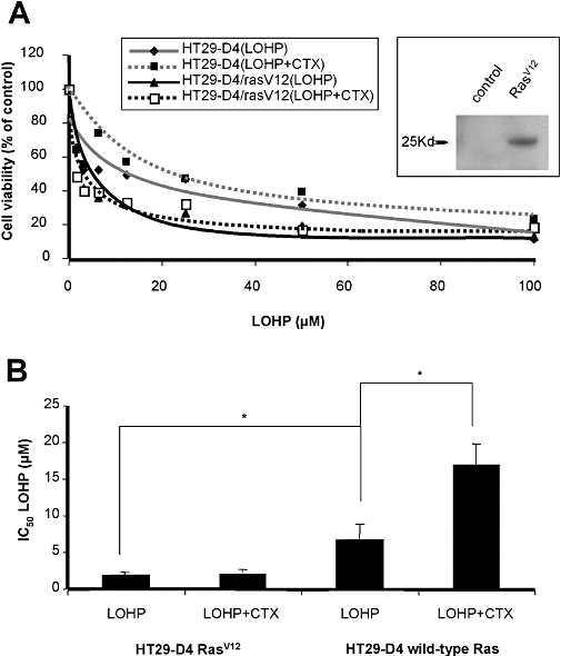
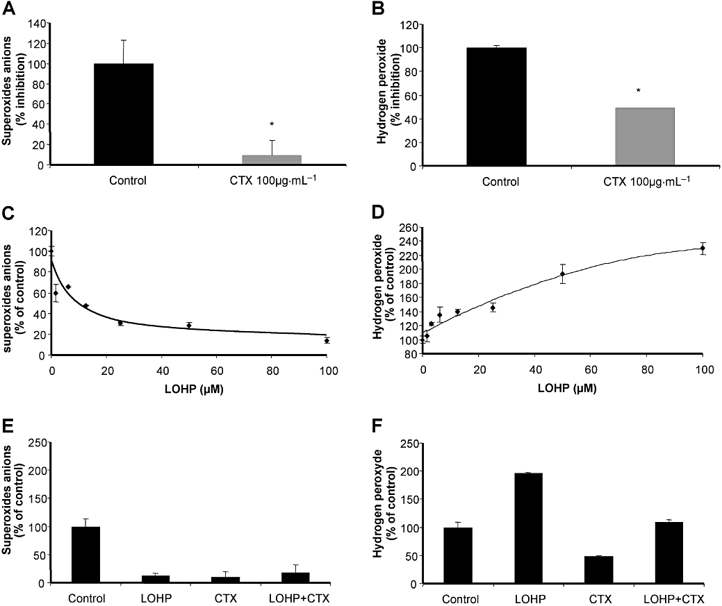
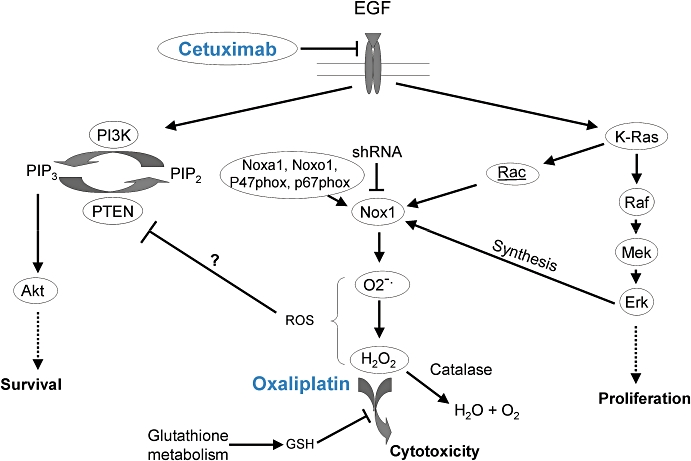
Key results: Combination of oxaliplatin and cetuximab was less cytotoxic than oxaliplatin alone in colorectal cells harbouring wild-type Ras and membrane expression of receptors for epidermal growth factor receptor (EGFR), such as HT29-D4 and Caco-2 cells. In contrast, cetuximab did not affect oxaliplatin efficiency in cells harbouring K-Ras(V12) mutation, irrespective of membrane EGFR expression (SW620 and SW480 cells). Transfection of HT29-D4 with K-Ras(V12) decreased oxaliplatin IC(50) and impaired cetuximab sensitivity, without affecting expression of membrane EGFR compared with HT29-D4 control. Oxaliplatin efficacy relies on endogenous production of H(2)O(2). Cetuximab inhibits H(2)O(2) production inhibiting the EGFR/Nox1 NADPH oxidase pathway. Oxaliplatin efficacy was impaired by short hairpin RNA for Nox1 and by catalase (H(2)O(2) scavenger).

Conclusions and implications: Cetuximab limited oxaliplatin efficiency by affecting the redox status of cancer cells through Nox1. Such combined therapy might be improved by controlling H(2)O(2) elimination.


Figures

Figure 1
Figure 1
(A) Epidermal growth factor receptor protein expression was detected by immunoblotting cell lysates from four colon cancer cell lines HT29-D4, Caco-2, SW480 and SW620. (B) Epidermal growth factor receptor cell surface expression was measured by flow cytometry. Cells (5 × 10⁵) were incubated with cetuximab as primary antibody and counterstained with an Alexa Fluor 488 goat anti-human IgG. All staining were done on ice for 45 min followed by three washes. For each cell line, a control without primary antibody was performed. (C) Detection by SNaPShot of K-Ras mutations on cell lines. Each peak corresponds to a specific extended primer. Wild type (WT) for HT29-D4 and Caco-2 (upper panel); K-Ras mutation for SW480 and SW620 (lower panel).
Figure 2
Figure 2
In vitro effects of a single agent, cetuximab (CTX) or oxaliplatin (LOHP), on a panel of human colorectal carcinoma cell lines. (A) Dose–response curves of cells treated with cetuximab alone at concentration ranging from 0.1 to 100 µg·mL−1 for 72 h on each cell lines using methylthiazoletetrazolium (MTT) assays. Results were presented as means ± SEM of three independent experiments. (B) Concentration–response curves of cells treated with oxaliplatin alone at concentrations ranging from 1 to 100 µM for 72 h using MTT assays. Data are expressed as mean ± SEM of three independent experiments. *P < 0.05. (C) IC50 for oxaliplatin combined with cetuximab in the panel of human colorectal carcinoma cell lines. Cells were treated with oxaliplatin at concentration ranging from 1 to 100 µM combined with a fixed cetuximab concentration of 100 µg·mL−1. Cetuximab was added 15 min before oxaliplatin. Growth inhibition was evaluated by using MTT assay. Data are expressed as mean ± SEM of three independent experiments.
Figure 3
Figure 3
(A) In vitro effects of oxaliplatin (LOHP) combined with cetuximab (CTX) in HT29-D4 control cells compared with HT29-D4 cells transfected with HA-tagged RasV12. Insert shows immunoblot for HA in transfected HT29-D4 cells. (B) IC50 values for oxaliplatin alone or combined with cetuximab on RasV12-transfected HT29-D4 cells compared with HT29-D4 control cells. *P < 0.05.
Figure 4
Figure 4
Production of O2 and H2O2 in HT29-D4 cells exposed to cetuximab (CTX) or oxaliplatin (LOHP). Production of O2 was determined by lucigenin and H2O2 production by DCFDA. Data from at least three independent experiments have been pooled. (A,B) Cetuximab significantly decreased O2 production and affected significantly the production of H2O2 compared with untreated cells (*P < 0.05). (C,D) Effects of oxaliplatin at concentrations ranging from 1 to 100 µM on O2 and H2O2 production. (E,F) Production of O2 and H2O2 in human colon carcinoma cells exposed to cetuximab, oxaliplatin or combination of the two drugs. Cells were treated with a fixed oxaliplatin concentration of 100 µM combined with a fixed cetuximab concentration of 100 µg·mL−1. Cetuximab was added 15 min before oxaliplatin. Cetuximab, oxaliplatin and the combination of both significantly decreased O2 production compared with untreated cells (P < 0.05). Oxaliplatin significantly increased H2O2 production, whereas cetuximab alone or combined with oxaliplatin decreased H2O2 production compared with untreated cells (P < 0.05).
Figure 5
Figure 5
(A) Nox1 protein expression detection by immunoblot in HT29-D4, Caco-2, SW480 and SW620 cell lines. (B) In vitro effect of oxaliplatin in HT29-D4 (upper panel) and Caco-2 cells (lower panel) transfected with Nox1 short hairpin RNA (shRNA) compared with control shRNA; Immunoblot for Nox1 in HT29-D4 cells transfected with indicated shRNA; blot is representative of three independent experiments. (C) In vitro effect of oxaliplatin in HT29-D4 with or without catalase used at non-cytotoxic concentrations.
Figure 6
Figure 6
Scheme of possible mechanisms underlying the antagonism between cetuximab and oxaliplatin, involving modulation of redox status. CTX, cetuximab; EGF, epidermal growth factor; GSH, glutathione; PI3K, phosphatidylinositol-3 kinase; PIP2, phosphatidylinositol bisphosphate; PIP3, phosphatidylinositol trisphosphate; PTEN, phosphatase and TENsin homologue, ROS, reactive oxygen species; shRNA, short hairpin RNA.

References

    1. Adachi Y, Shibai Y, Mitsushita J, Shang WH, Hirose K, Kamata T. Oncogenic Ras upregulates NADPH oxidase 1 gene expression through MEK-ERK-dependent phosphorylation of GATA-6. Oncogene. 2008;27:4921–4932. - PubMed
    1. Alexander SP, Mathie A, Peters JA. Guide to Receptors and Channels (GRAC) Br J Pharmacol. (3rd) 2008;153(Suppl 2):S1–209. - PMC - PubMed
    1. Alexandre J, Nicco C, Chereau C, Laurent A, Weill B, Goldwasser F, et al. Improvement of the therapeutic index of anticancer drugs by the superoxide dismutase mimic mangafodipir. J Natl Cancer Inst. 2006;98:236–244. - PubMed
    1. Balin-Gauthier D, Delord JP, Rochaix P, Mallard V, Thomas F, Hennebelle I, et al. In vivo and in vitro antitumor activity of oxaliplatin in combination with cetuximab in human colorectal tumor cell lines expressing different level of EGFR. Cancer Chemother Pharmacol. 2006;57:709–718. - PubMed
    1. Banfi B, Schrenzel J, Nusse O, Lew DP, Ligeti E, Krause KH, et al. A novel H(+) conductance in eosinophils: unique characteristics and absence in chronic granulomatous disease. J Exp Med. 1999;190:183–194. - PMC - PubMed

Publication types

MeSH terms