

Changes in collagen fibril network organization and proteoglycan distribution in equine articular cartilage during maturation and growth
- PMID: 19732210
- PMCID: PMC2780575
- DOI: 10.1111/j.1469-7580.2009.01140.x
Changes in collagen fibril network organization and proteoglycan distribution in equine articular cartilage during maturation and growth
Abstract
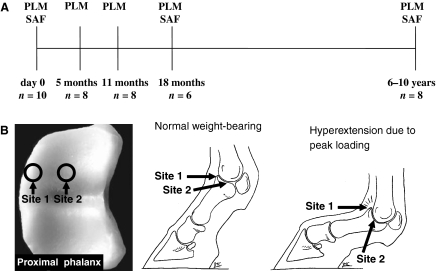
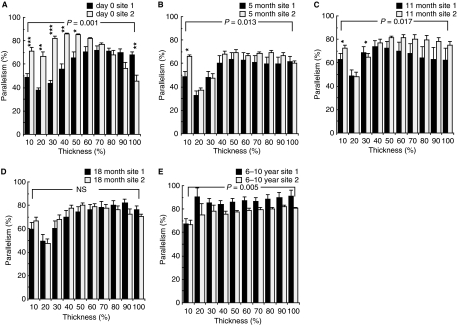
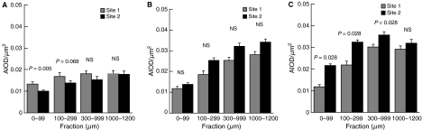
The aim of this study was to record growth-related changes in collagen network organization and proteoglycan distribution in intermittently peak-loaded and continuously lower-level-loaded articular cartilage. Cartilage from the proximal phalangeal bone of the equine metacarpophalangeal joint at birth, at 5, 11 and 18 months, and at 6-10 years of age was collected from two sites. Site 1, at the joint margin, is unloaded at slow gaits but is subjected to high-intensity loading during athletic activity; site 2 is a continuously but less intensively loaded site in the centre of the joint. The degree of collagen parallelism was determined with quantitative polarized light microscopy and the parallelism index for collagen fibrils was computed from the cartilage surface to the osteochondral junction. Concurrent changes in the proteoglycan distribution were quantified with digital densitometry. We found that the parallelism index increased significantly with age (up to 90%). At birth, site 2 exhibited a more organized collagen network than site 1. In adult horses this situation was reversed. The superficial and intermediate zones exhibited the greatest reorganization of collagen. Site 1 had a higher proteoglycan content than site 2 at birth but here too the situation was reversed in adult horses. We conclude that large changes in joint loading during growth and maturation in the period from birth to adulthood profoundly affect the architecture of the collagen network in equine cartilage. In addition, the distribution and content of proteoglycans are modified significantly by altered joint use. Intermittent peak-loading with shear seems to induce higher collagen parallelism and a lower proteoglycan content in cartilage than more constant weight-bearing. Therefore, we hypothesize that the formation of mature articular cartilage with a highly parallel collagen network and relatively low proteoglycan content in the peak-loaded area of a joint is needed to withstand intermittent stress and shear, whereas a constantly weight-bearing joint area benefits from lower collagen parallelism and a higher proteoglycan content.
Figures
Similar articles
-
Influence of exercise and joint topography on depth-related spatial distribution of proteoglycan and collagen content in immature equine articular cartilage.Equine Vet J. 2009 Jul;41(6):557-63. doi: 10.2746/042516409x424162. Equine Vet J. 2009. PMID: 19803051 Clinical Trial.
-
Changes in spatial collagen content and collagen network architecture in porcine articular cartilage during growth and maturation.Osteoarthritis Cartilage. 2009 Apr;17(4):448-55. doi: 10.1016/j.joca.2008.09.004. Epub 2008 Oct 11. Osteoarthritis Cartilage. 2009. PMID: 18849174
-
Topographical mapping of biochemical properties of articular cartilage in the equine fetlock joint.Equine Vet J. 2000 Jan;32(1):19-26. doi: 10.2746/042516400777612062. Equine Vet J. 2000. PMID: 10661380
-
Quantitative magnetic resonance imaging of the articular cartilage of the knee joint.Magn Reson Imaging Clin N Am. 2014 Nov;22(4):649-69. doi: 10.1016/j.mric.2014.07.005. Epub 2014 Nov 1. Magn Reson Imaging Clin N Am. 2014. PMID: 25442027 Review.
-
The Effect of Aging and Mechanical Loading on the Metabolism of Articular Cartilage.J Rheumatol. 2017 Apr;44(4):410-417. doi: 10.3899/jrheum.160226. Epub 2017 Mar 1. J Rheumatol. 2017. PMID: 28250141 Review.
Cited by
-
Systematic Comparison of Biomaterials-Based Strategies for Osteochondral and Chondral Repair in Large Animal Models.Adv Healthc Mater. 2021 Oct;10(20):e2100878. doi: 10.1002/adhm.202100878. Epub 2021 Aug 18. Adv Healthc Mater. 2021. PMID: 34405587 Free PMC article.
-
Composition, structure and tensile biomechanical properties of equine articular cartilage during growth and maturation.Sci Rep. 2018 Jul 27;8(1):11357. doi: 10.1038/s41598-018-29655-5. Sci Rep. 2018. PMID: 30054498 Free PMC article.
-
Effects of serum and compressive loading on the cartilage matrix synthesis and spatiotemporal deposition around chondrocytes in 3D culture.Tissue Eng Part A. 2013 May;19(9-10):1199-208. doi: 10.1089/ten.tea.2012.0559. Epub 2013 Feb 14. Tissue Eng Part A. 2013. PMID: 23410025 Free PMC article.
-
Slope-based segmentation of articular cartilage using polarization-sensitive optical coherence tomography phase retardation image.J Biomed Opt. 2019 Mar;24(3):1-14. doi: 10.1117/1.JBO.24.3.036006. J Biomed Opt. 2019. PMID: 30873765 Free PMC article.
-
Cartilage canals in the distal intermediate ridge of the tibia of fetuses and foals are surrounded by different types of collagen.J Anat. 2017 Oct;231(4):615-625. doi: 10.1111/joa.12650. Epub 2017 Jun 15. J Anat. 2017. PMID: 28620929 Free PMC article.
References
-
- Arokoski JPA, Hyttinen MM, Lapvetalainen T, et al. Decreased birefringence of the superficial zone collagen network in the canine knee (stifle) articular cartilage after long distance running training, detected by quantitative polarised light microscopy. Ann Rheum Dis. 1996;55:253–264. - PMC - PubMed
-
- Aspden RM, Hukins DWL. Collagen organization in articular cartilage, determined by X-ray diffraction, and its relationship to tissue function. Proc R Soc Lond Biol Sci. 1981;212:299–304. - PubMed
-
- Bader DL, Kempson GE, Egan J, et al. The effects of selective matrix degradation on the short-term compressive properties of adult human articular cartilage. Biochim Biophys Acta – Gen Sub. 1992;1116:147–154. - PubMed
-
- Benninghoff A. Form und Bau der Gelenkknorpel in ihren Beziehungen zur Funktion. Zweiter teil: Der Aufnau des Gelenkknorpels in seinen Beziehungen zur Funktion. Z Zelforsch Mikrosk Anat. 1925;2:783–862.
-
- Brama PA, Tekoppele JM, Bank RA, et al. Influence of different exercise levels and age on the biochemical characteristics of immature equine articular cartilage. Equine Vet J Suppl. 1999a;31:55–61. - PubMed
Publication types
MeSH terms
Substances
LinkOut - more resources
Full Text Sources
