

The molecular and genetic mechanisms of neocortex development
- PMID: 19732610
- PMCID: PMC2771632
- DOI: 10.1016/j.clp.2009.06.008
The molecular and genetic mechanisms of neocortex development
Abstract
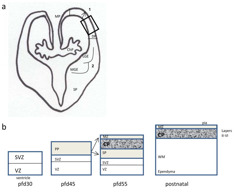
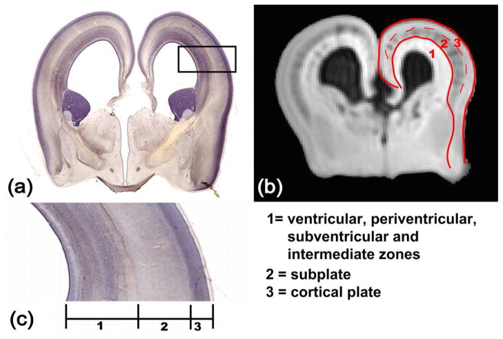
This article reviews key recent findings in the field of human cortical development. This development is divided into three major time-dependent phases: neural proliferation of inhibitory and excitatory neurons in spatially distinct regions, migration through multiple cellular boundaries, and maturation through morphologic changes that result in the elaboration of dendrites and axons and that establish the multitude of cellular contacts that underlie neuronal processing. Many of the neurocognitive disorders treated in the clinic can trace their origin to a disorder in one or more of these key steps. Along with this update, work is highlighted that offers a glimpse at the future of therapy for developmental brain disorders that can result from disorders of these cellular events.
Figures
References
-
- Chitnis AB. Control of neurogenesis--lessons from frogs, fish and flies. Curr Opin Neurobiol. 1999;9:18–25. - PubMed
-
- O’Rahilly R, Müller F. The embryonic human brain: an atlas of developmental stages. 3. Hoboken, NJ: Wiley-Liss; 2006.
-
- Bystron I, Blakemore C, Rakic P. Development of the human cerebral cortex: Boulder Committee revisited. Nat Rev Neurosci. 2008;9:110–22. - PubMed
-
- Anthony TE, Klein C, Fishell G, et al. Radial glia serve as neuronal progenitors in all regions of the central nervous system. Neuron. 2004;41:881–90. - PubMed
Publication types
MeSH terms
Grants and funding
LinkOut - more resources
Full Text Sources
Medical
