. 2009 Sep 8;16(3):183-94.
doi: 10.1016/j.ccr.2009.06.017.

Polarization of tumor-associated neutrophil phenotype by TGF-beta: "N1" versus "N2" TAN

Affiliations

Polarization of tumor-associated neutrophil phenotype by TGF-beta: "N1" versus "N2" TAN

Zvi G Fridlender et al. Cancer Cell. .

Abstract

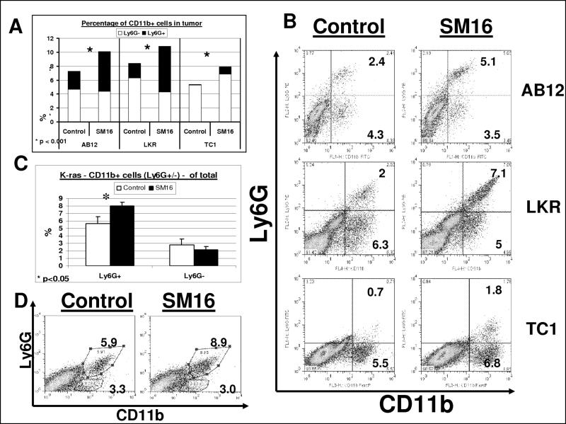
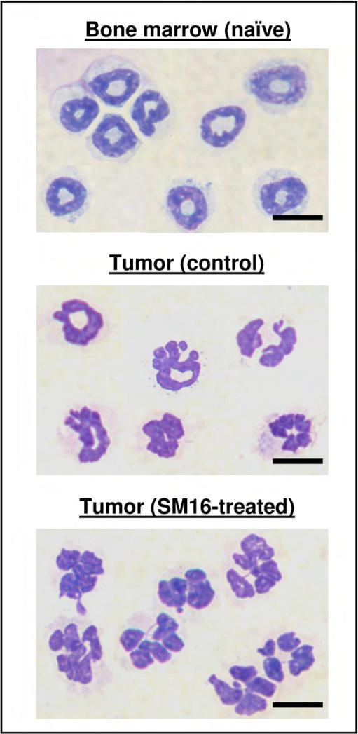
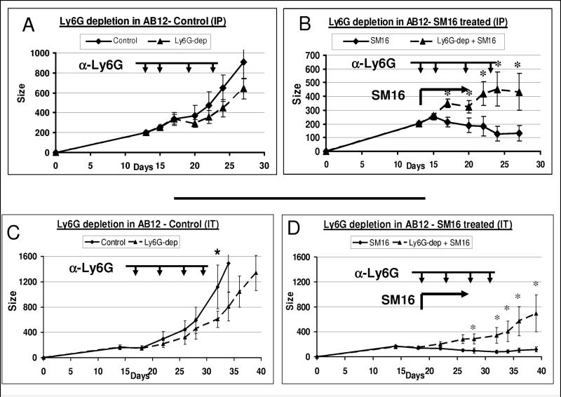
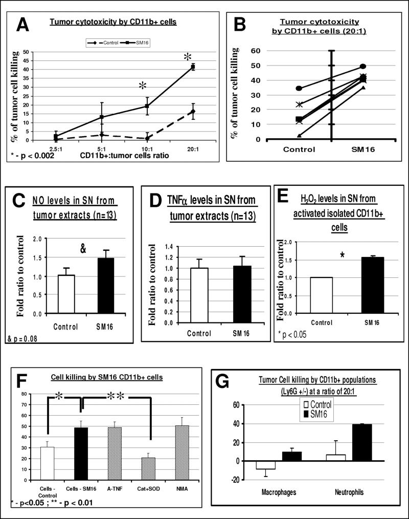
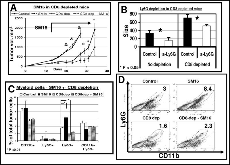
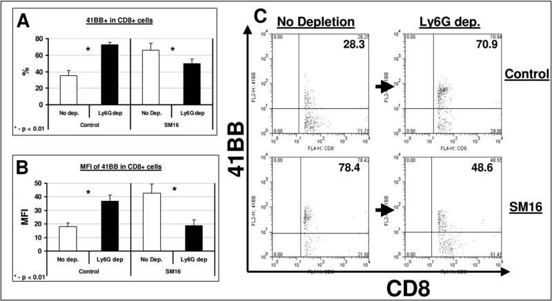
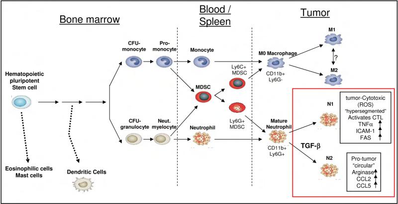
TGF-beta blockade significantly slows tumor growth through many mechanisms, including activation of CD8(+) T cells and macrophages. Here, we show that TGF-beta blockade also increases neutrophil-attracting chemokines, resulting in an influx of CD11b(+)/Ly6G(+) tumor-associated neutrophils (TANs) that are hypersegmented, more cytotoxic to tumor cells, and express higher levels of proinflammatory cytokines. Accordingly, following TGF-beta blockade, depletion of these neutrophils significantly blunts antitumor effects of treatment and reduces CD8(+) T cell activation. In contrast, in control tumors, neutrophil depletion decreases tumor growth and results in more activated CD8(+) T cells intratumorally. Together, these data suggest that TGF-beta within the tumor microenvironment induces a population of TAN with a protumor phenotype. TGF-beta blockade results in the recruitment and activation of TANs with an antitumor phenotype.


Figures

Figure 1
Figure 1. SM16 causes an influx of CD11b+ Ly6G+ granulocytic cells into tumors
Panels A-B. Flow cytometry was performed on digested tumors from animals treated for one week with control chow (left columns) or SM16 chow (right columns), in each of the three flank tumors – AB12 (n=26), LKR (n=5) and TC-1 (n=9−10). Panel A summarizes the percentage of CD11b+ cells out of all tumor cells in the three cell lines, in both groups (total). This is divided to Ly6G cells (bottom section of each bar- white), and Ly6G+ (granulocytic) cells (top section of each bar- black). *=p<0.001. Panel B shows representative FACS tracings of CD11b versus Ly6G expression in each of the lines. The number in each quadrant is the percentage of the total tumor cells. Panels C-D. Flow cytometry was performed on digested lungs with orthotopic tumors from mice with the conditionally expressed, K-rasG12D allele, treated for one week with control (white) or SM16 (black) chow (n=5 per group). Panel C summarizes the percentage of the different CD11b+ cells out of all lung cells ± SEM – Ly6G+ (granulocytic) cells (left) and Ly6G cells (right). *=p<0.05. Panel D shows representative FACS tracings of CD11b versus Ly6G expression in the two groups. The number in each quadrant is the percentage of total lung cells.
Figure 2
Figure 2. The morphology of TAN in control and SM16-treated mice compared to bone-marrow neutrophils
Photomicrograph slides from bone marrow neutrophils (top panel) and from previously sorted CD11b+/Ly6G+ cells from control (middle panel) or SM16-treated mice (bottom panel). Scale bars 10 μm.
Figure 3
Figure 3. Effect of neutrophil depletion on tumor growth and on tumor response to SM16
Panels A-B (systemic depletion). Mice (n = 6−8 for each subgroup) bearing large AB12 tumors, were treated with either control (Panel A) or SM16 chow (panel B), starting at day 13. One group on each diet was injected with either 100 μg of the anti-Ly6G monoclonal antibody 1A8 intra-peritoneally (IP) (arrowheads) every 3−5 days during the experiment (triangles, with dashed lines) or a control IgG antibody at the same schedule and dose (diamonds with solid lines). Panel A compares mean tumor size ± SEM with or without Ly6G depletion (Ly6G-dep) in mice treated with control chow. Panel B compares mean tumor size ± SEM with or without Ly6G depletion in mice treated with SM16 chow. Groups were compared using ANOVA. *=p<0.05. Panels C-D (intra-tumoral depletion). The experiment was repeated again with injection of 30 μg of the anti-Ly6G monoclonal antibody 1A8 or control IgG given intratumorally (IT, arrowheads). Groups were compared using ANOVA. *=p<0.05.
Figure 4
Figure 4. SM16 CD11b+ cell cytotoxicity is primarily due to Activated Neutrophils, via an oxygen radical-dependent mechanism
Panels A-B. AB12 tumors (n=5−7 for each group) from control and SM16-treated animals were treated for 7 days and then digested and pooled. Isolated CD11b+ cells were co-cultured with AB12-luciferase cells at different ratios of effector cells (CD11b+) to tumor cells. At 24 hours, the percentage of tumor cells killed was calculated. Panel A summarizes the percentage of tumor killing ± SEM at each ratio of co-culture (n=4−6) *=p<0.002. In panel B, the individual data from 6 separate experiments with co-culture at a ratio of 20 effector cells to 1 tumor cell is shown. Panels C-D. Pieces of harvested tumors from control and SM16-treated mice were cultured in medium for 24 hours, and the secretion of NO (panel C) and TNFα (Panel D) per mg of tissue was evaluated. The bars represent mean ± SEM, &-p=0.08. Panel E. Isolated CD11b+ cells were cultured in wells and activated with PMA, followed by evaluation of the release of H2O2. The bars represent mean ± SEM, *=p<0.05. Panel F. CD11b+ cells were co-cultured with AB12-Luc cells as above (panels A-B) and different inhibitors were added. The bars represent mean ± SEM. *=p<0.05, **=p<0.01. Panel G. CD11b+ cells were sorted using anti-Ly6G antibody to neutrophils (Ly6G+) and macrophages (Ly6G). Each of the cell subtypes were co-cultured with tumor cells at a ratio of 20 effector cells to 1 tumor cell, and cytotoxicity was evaluated as above. The bars represent mean ± SEM.
Figure 5
Figure 5. CD8+ cell depletion blocks all of the SM16 clinical effect and reduces influx of neutrophils to the tumors
Panel A. - Mice (n = 5−6 for each group) bearing large AB12 tumors were treated in one of four ways: 1) control chow (diamonds- control); 2) SM16 chow starting at day 13 and throughout the experiment (squares - SM16); 3) control chow, and injected with 300 μg of an anti-CD8 monoclonal antibody IP twice per week starting two days prior to tumor injection (triangles – CD8 dep.); and 4) SM16 chow and depletion of CD8+ cells (crosses – CD8-dep - SM16). Control and SM16 groups were treated with an IP control IgG antibody. The bars represent mean ± SEM. *=p<0.05, **=p<0.01 - control vs SM16; &=p<0.05 control vs. SM16 (both with CD8 depletion). Panel B. - Mice were injected with either 300 μg of an anti-CD8 monoclonal antibody IP twice per week starting two days prior to tumor injection (right – CD8 depleted) or control IgG (left – no depletion). When tumors reach a size of approximately 100mm3, Each of these two groups (n = 10−12 for each group) were divided to two subgroups, treated for 2 weeks with either a control IgG antibody (Control, black) or 100 μg of an anti-Ly6G monoclonal antibody i.t. twice per week (α-Ly6G, white), followed by tumor measurements. The bars represent mean size ± SEM of each group. Although CD8 depletion accelerated tumor growth, the reduction in tumor growth following Ly6G depletion was maintained in this group (right panel). Differences in both groups were significant (*=p<0.05). Panels C-D. - Mice (n = 4−5 for each group) bearing large AB12 tumors were treated in one of four ways as above. Seven days after starting treatment with SM16, flow cytometry of the tumors was performed. Panel C summarizes the percentage of CD11b+, Ly6G+, Ly6C+ and CD11b+ Ly6G cells in the four groups out of the total number of tumor cells ± SEM, *=p<0.05. Panel D shows representative FACS tracings of CD11b versus Ly6G expression in each of the four treatment groups. The number in each panel is the percentage of the total tumor cells.
Figure 6
Figure 6. Neutrophil depletion increases CD8+ T-cell activity in untreated mice, but reduces CD8+ T-cell activity in SM16-treated mice
Mice (n = 5 for each group) bearing large AB12 tumors were treated in one of four ways: control chow (control); SM16 chow (SM16); control chow and injected with 100 μg of the anti-Ly6G monoclonal antibody 1A8 IP twice per week (Ly6G-dep); SM16 chow and depletion of neutrophils (Ly6G-dep + SM16). The groups not treated with Ly6G depletion (Control and SM16) were treated with a control IgG antibody at the same schedule and dose. Seven days after starting SM16 or control chow, multi-color flow cytometry of tumors was performed. Activation of the CD8+ T-cells was measured using the activity marker 4−1BB (CD137). T cell activation was compared with and without neutrophil-depletion in the control-chow-treated tumor bearing mice (left columns) and SM16-treated tumor-bearing mice (right columns) tumors. Panel A summarizes the percentage of 4−1BB+ out of total intra-tumoral CD8+ T-cells ± SEM. Panel B summarizes the Mean Fluorescent Intensity (MFI) of 4−1BB in the intra-tumoral CD8+ T-cells ± SEM, *=P<0.01. Panel C shows representative FACS tracings of CD8 versus 4−1BB expression in each of the four treatment groups. The number in the upper right quadrant is the percentage of 4−1BB+ cells out of total CD8+ cells.
Figure 7
Figure 7. The origin and differentiation of myeloid-derived tumor associated cells (based on (Murdoch et al., 2008) and (Gabrilovich and Nagaraj, 2009))
Myeloid-derived tumor associated cells originate from a common pluripotent stem cell, but separate early to monocytic and granulocytic lineages, eventually infiltrating tumors. As we show in the current study, TAN can polarize to either anti-tumor N1 TAN or pro-tumor N2 TAN, with TGF-β being an important effector in that polarization. The characteristics of the polarized TAN, as presented in the current study, are framed in the bottom right part of the figure.

Comment in

References

    1. Allavena P, Sica A, Garlanda C, Mantovani A. The Yin-Yang of tumor-associated macrophages in neoplastic progression and immune surveillance. Immunological Reviews. 2008;222:155–161. - PubMed
    1. Allen SS, Mackie JT, Russell K, Jeevan A, Skwor TA, McMurray DN. Altered inflammatory responses following transforming growth factor-[beta] neutralization in experimental guinea pig tuberculous pleurisy. Tuberculosis. 2008;88:430–436. - PubMed
    1. Appelberg R. Mycobacterial infection primes T cells and macrophages for enhanced recruitment of neutrophils. J Leukoc Biol. 1992;51:472–477. - PubMed
    1. Aruga A, Aruga E, Cameron MJ, Chang AE. Different cytokine profiles released by CD4+ and CD8+ tumor-draining lymph node cells involved in mediating tumor regression. J Leukoc Biol. 1997;61:507–516. - PubMed
    1. Balkwill F, Coussens LM. Cancer: An inflammatory link. Nature. 2004;431:405–406. - PubMed

Publication types

MeSH terms