. 2009 Sep 8;16(3):259-66.
doi: 10.1016/j.ccr.2009.07.016.

FKBP51 affects cancer cell response to chemotherapy by negatively regulating Akt

Affiliations

FKBP51 affects cancer cell response to chemotherapy by negatively regulating Akt

Huadong Pei et al. Cancer Cell. .

Abstract

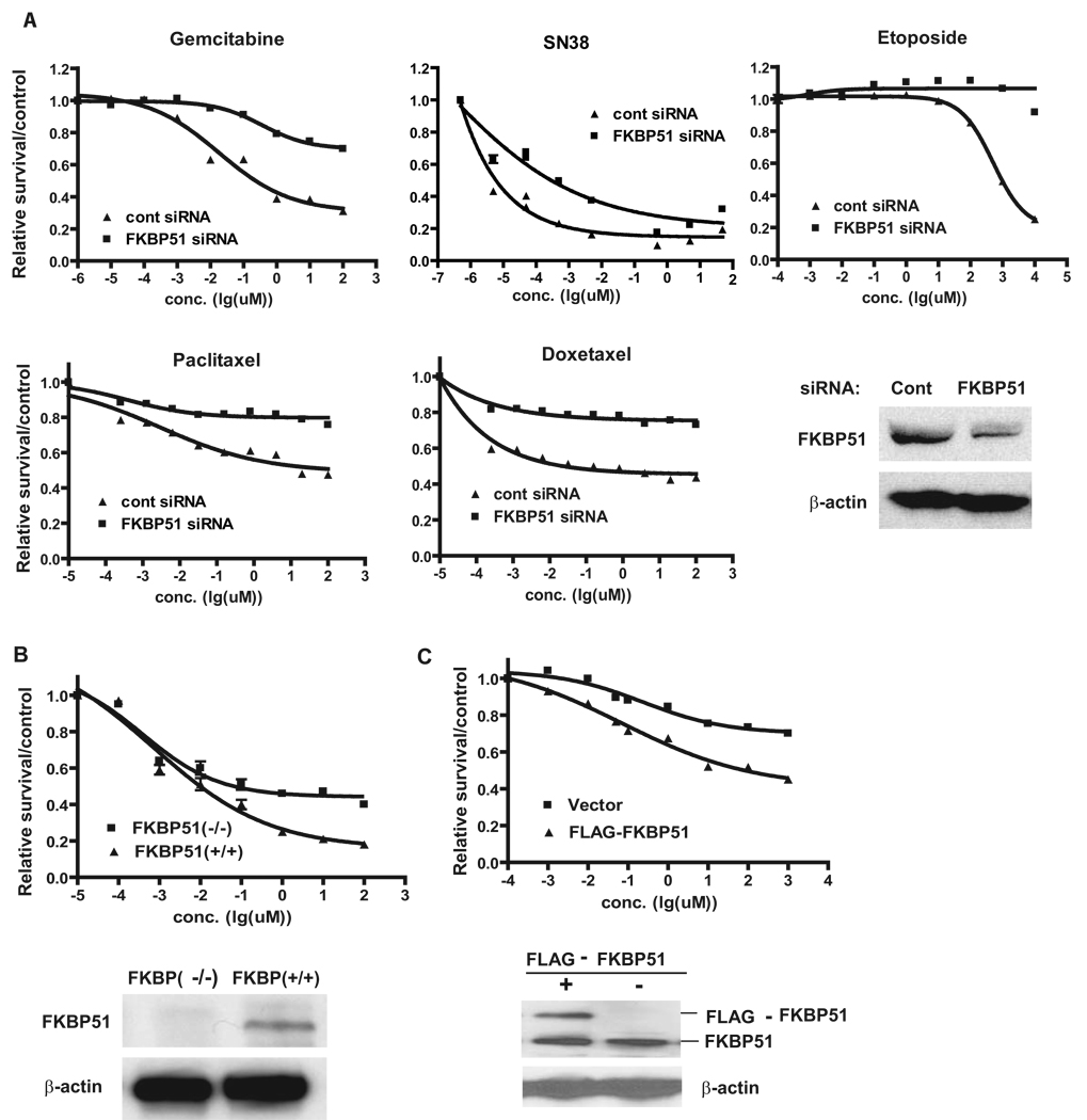
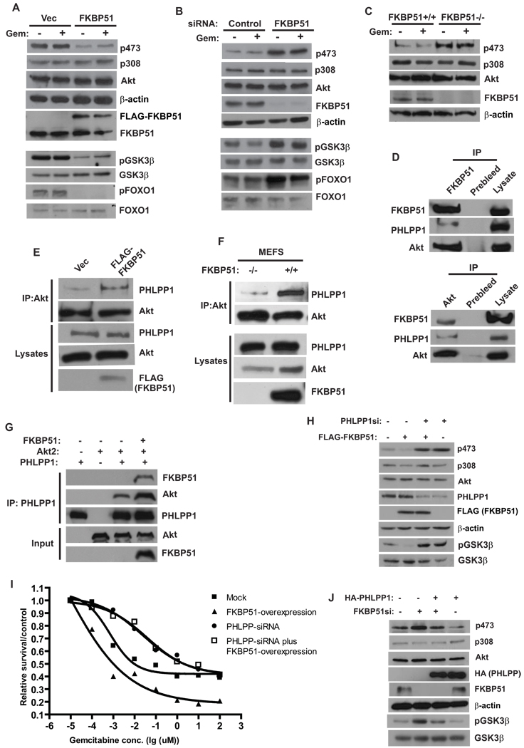
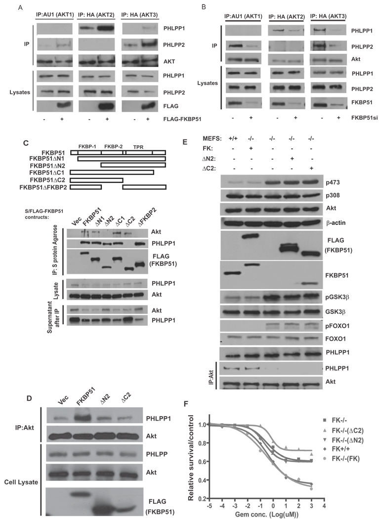
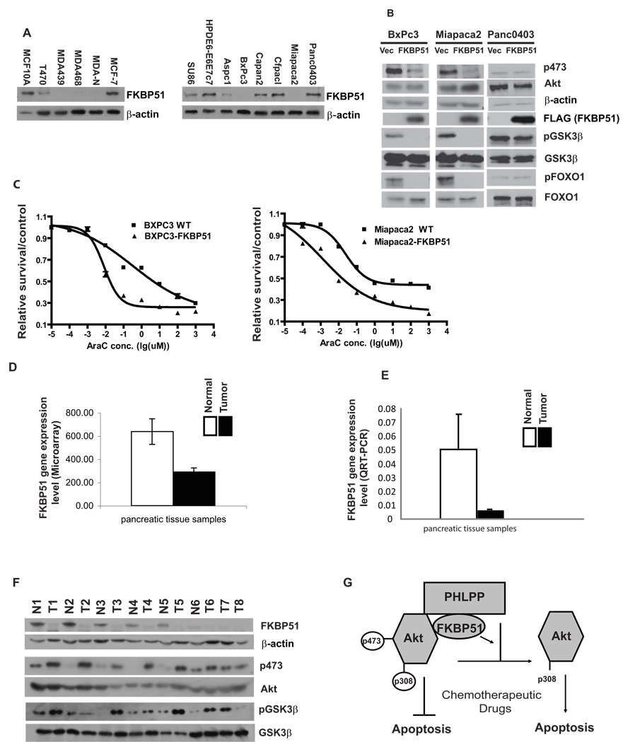
Akt is a central regulator of cell growth. Its activity can be negatively regulated by the phosphatase PHLPP that specifically dephosphorylates the hydrophobic motif of Akt (Ser473 in Akt1). However, how PHLPP is targeted to Akt is not clear. Here we show that FKBP51 (FK506-binding protein 51) acts as a scaffolding protein for Akt and PHLPP and promotes dephosphorylation of Akt. Furthermore, FKBP51 is downregulated in pancreatic cancer tissue samples and several cancer cell lines. Decreased FKBP51 expression in cancer cells results in hyperphosphorylation of Akt and decreased cell death following genotoxic stress. Overall, our findings identify FKBP51 as a negative regulator of the Akt pathway, with potentially important implications for cancer etiology and response to chemotherapy.


Figures

Figure 1
Figure 1. FKBP51 regulates cellular response to genotoxic stress
(A). SU86 cells were transfected with control or FKBP51 siRNA and then treated with the indicated drugs. Cell survival was determined as described in the Methods. (B). FKBP51+/+ or FKBP51−/− cells were treated with gemcitabine, and cell survival was determined as in (A). (C). SU86 cells were transfected with vector or constructs encoding FLAG-FKBP51. Transfected cells were treated with gemcitabine, and cell survival was determined as in (A). Points, mean values for three independent experiments; Error bars, +/− SEM.
Figure 2
Figure 2. FKBP51 regulates Akt phosphorylation at Ser473 by promoting Akt-PHLPP interaction
(A). SU86 cells were transfected for 48 hr with indicated constructs. Cells were treated with DMSO or gemcitabine (Gem, 20 nM, 12 hrs), and the phosphorylation of Akt, FOXO and GSK-3β in cell lysates was detected by Western Blot. (B). SU86 cells were transfected with indicated siRNA. Cells were then treated and harvested as in (A). (C). The phosphorylation of Akt in cell lysates from FKBP51+/+ or FKBP51−/− cells was examined. (D). The coimmunoprecipitation of Akt, PHLPP and FKBP51 was examined. (E). 293T cells were transfected with indicated constructs, and the interaction between Akt and PHLPP was examined. (F). The PHLPP-Akt interaction was examined in FKBP51+/+ and FKBP51−/− cells. (G). Purified recombinant Akt, PHLPP1 and FKBP51 were incubated in vitro as indicated. The Akt-PHLPP interaction was then examined by coimmunoprecipitation. (H–I). SU86 cells were transfected with FLAG-FKBP51 and/or PHLPP siRNA as indicated. The phosphorylation of Akt, GSK-3β, as well as sensitivity to gemcitabine were then examined. Points, mean values for three independent experiments; Error bars, +/− SEM. (J). SU86 cells were transfected with HA-PHLPP and/or FKBP51 siRNA as indicated. The phosphorylation of Akt and GSK-3β was then examined.
Figure 3
Figure 3. FKBP51 scaffolding function regulates Akt phosphorylation and cell survival
(A). 293T cells or 293T cells stably transfected with FKBP51 were transfected with AKT isoforms (lane 1,2, AU1-AKT1; lane 3,4, HA-AKT2, lane5,6 HA-AKT3). Cells were lyzed, and lysates were subjected to immunoprecipitation with indicated antibodies. PHLPP1, PHLPP2 and AKT in the immunoprecipitates or cell lysates were detected by immunoblotting. (B). SU86 cells were transfected with control or FKBP51 siRNA together with AKT isoforms. The interaction between Akt isoforms and PHLPP isoforms was then examined as in A. (C). 293T cells were transfected with different S/FLAG-tagged FKBP51 truncated mutants. Lysates from transfected cells were subjected to immunoprecipitation with S protein agarose, PHLPP1 and Akt in the immunoprecipitates were then detected by immunoblotting. (D). 293T cells were transfected with WT FKBP51 or FKBP51 truncation mutations. Transfected cells were then lyzed, and lysates were subjected to immunoprecipitation with anti-Akt antibodies. PHLPP1 and Akt in the immunoprecipitates or cell lysates were detected by immunoblotting. (E–F). FKBP51+/+, FKBP51−/− or FKBP51−/− MEFs stably expressing WT or mutant FKBP51 were used to examine Akt, GSK-3β and FOXO1 phosphorylation as well as the Akt-PHLPP interaction (E). cells were examined for gemcitabine sensitivity using the MTS assay (F). Points, mean values for three independent experiments; Error bars, +/− SEM.
Figure 4
Figure 4. Loss of FKBP51 expression in cancer cells and tissues
(A). Cell lysates from pancreatic and breast cancer cell lines were blotted with FKBP51 antibodies. Lysates from normal breast (MCF10A) and pancreatic (HPDE6-E6E7c7) epithelial cells were used as controls. (B–C). Miapaca2, BxPC3 or Panc0403 cells were reconstituted with FKBP51, and Akt phosphorylation and sensitivity to genotoxic stress were then determined. Points, mean values for three independent experiments; Error bars, +/− SEM. (D, E). FKBP51 gene expression in tumor and normal pancreatic tissues was determined using microarray analysis (D, 19 normal pancreatic and 36 tumor tissue samples, p= 0.0092) or real-time QRT-PCR (E, 25 pancreatic cancer samples and 12 normal pancreatic tissues, p=0.001). Error bars, +/− SEM. (F). Western blot of lysates from a subset of tumor and normal tissues. T: Tumor; N: Normal pancreatic tissue; 1–8, patient No. (G). A model illustrates how FKBP51 regulates cell survival through the Akt pathway.

Comment in

References

    1. Alessi DR, Andjelkovic M, Caudwell B, Cron P, Morrice N, Cohen P, Hemmings BA. Mechanism of activation of protein kinase B by insulin and IGF-1. Embo J. 1996;15:6541–6551. - PMC - PubMed
    1. Alessi DR, James SR, Downes CP, Holmes AB, Gaffney PR, Reese CB, Cohen P. Characterization of a 3-phosphoinositide-dependent protein kinase which phosphorylates and activates protein kinase Balpha. Curr Biol. 1997;7:261–269. - PubMed
    1. Bai X, Ma D, Liu A, Shen X, Wang QJ, Liu Y, Jiang Y. Rheb activates mTOR by antagonizing its endogenous inhibitor, FKBP38. Science. 2007;318:977–980. - PubMed
    1. Barent RL, Nair SC, Carr DC, Ruan Y, Rimerman RA, Fulton J, Zhang Y, Smith DF. Analysis of FKBP51/FKBP52 chimeras and mutants for Hsp90 binding and association with progesterone receptor complexes. Mol Endocrinol. 1998;12:342–354. - PubMed
    1. Baughman G, Wiederrecht GJ, Campbell NF, Martin MM, Bourgeois S. FKBP51, a novel T-cell-specific immunophilin capable of calcineurin inhibition. Mol Cell Biol. 1995;15:4395–4402. - PMC - PubMed

Publication types

MeSH terms

Associated data