. 2009 Sep;85(3):328-37.
doi: 10.1016/j.ajhg.2009.07.017.

Mutations in LOXHD1, an evolutionarily conserved stereociliary protein, disrupt hair cell function in mice and cause progressive hearing loss in humans

Affiliations

Mutations in LOXHD1, an evolutionarily conserved stereociliary protein, disrupt hair cell function in mice and cause progressive hearing loss in humans

Nicolas Grillet et al. Am J Hum Genet. 2009 Sep.

Abstract

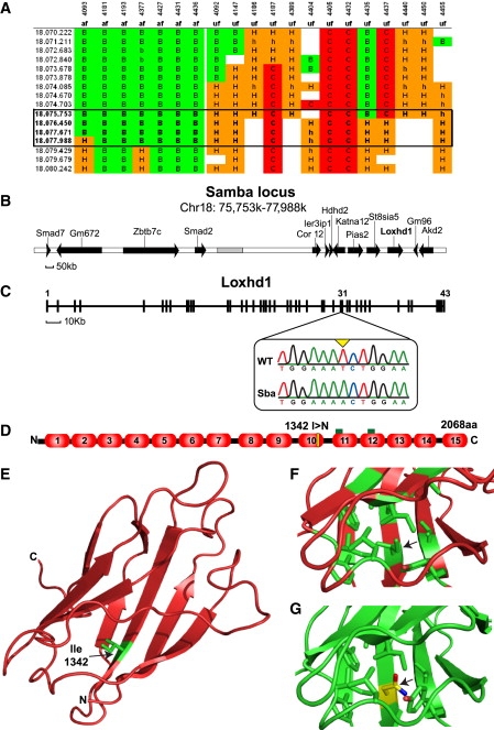
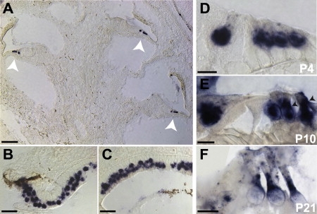
Hearing loss is the most common form of sensory impairment in humans and is frequently progressive in nature. Here we link a previously uncharacterized gene to hearing impairment in mice and humans. We show that hearing loss in the ethylnitrosourea (ENU)-induced samba mouse line is caused by a mutation in Loxhd1. LOXHD1 consists entirely of PLAT (polycystin/lipoxygenase/alpha-toxin) domains and is expressed along the membrane of mature hair cell stereocilia. Stereociliary development is unaffected in samba mice, but hair cell function is perturbed and hair cells eventually degenerate. Based on the studies in mice, we screened DNA from human families segregating deafness and identified a mutation in LOXHD1, which causes DFNB77, a progressive form of autosomal-recessive nonsyndromic hearing loss (ARNSHL). LOXHD1, MYO3a, and PJVK are the only human genes to date linked to progressive ARNSHL. These three genes are required for hair cell function, suggesting that age-dependent hair cell failure is a common mechanism for progressive ARNSHL.


Figures

Figure 1
Figure 1
Auditory Defects in samba Mice at 3 Weeks of Age (A) Representative click-evoked ABRs for wild-type (C57Bl6/J) mice and homozygous samba mutants at different sound intensities. ABR peaks I-IV are indicated. samba mutants showed no response even at the highest sound intensity (90 dB). (B) Average auditory thresholds in 3-week-old mice (C57Bl/6J, n = 5; Sba+/−, n = 4; Sba−/−, n = 6; mean ± SD; ∗∗∗p < 0.001, Student's t test). (C) Representative DPOAE response spectra from a 3-week-old wild-type and homozygous samba mouse at a single stimulus condition (median primary frequency = 16 kHz). The cubic distortion product (2f1-f2) was absent in the mutants (black arrow). (D) DPOAE thresholds in 3-week-old samba mutants were elevated at all frequencies analyzed (wild-type, n = 4; samba, n = 6; mean ± SD). Primary frequencies were maintained at an f2/f1 ratio of 1.22 and L1 was equal to L2.
Figure 2
Figure 2
samba Mice Carry a Missense Mutation in Loxhd1 (A) DNA from affected (af) and unaffected (uf) mice was analyzed for SNP markers on chromosome 18, which are listed in the first column (the megabase position 70.222–80.242 is indicated). The mouse identification number and phenotypes are shown in the two top rows. The genotype for each marker is given as: B, homozygous C57Bl/6J; C, homozygous 129; H, heterozygous. The chromosomal interval that segregates with the mutant phenotype is highlighted in the box. (B) The annotated genes in the affected interval in samba mice on chromosome 18 are indicated. (C) Predicted exon/intron structure of the Loxhd1 gene. Exons are shown as black squares. Several exons are numbered. The sequence chromatogram shows the mutation in exon 31. (D) The LOXHD1 protein is predicted to consist of 15 PLAT domains. The samba mutation (1342 I>N) is indicated by a yellow bar, and the locations of the immunization peptides by green bars. (E) Diagram of the PLAT 10 wild-type model. The cartoon is based on the C-alpha positions of the PLAT 10 model, which was derived from the PLAT domain of the allene oxide synthase-lipoxygenase coordinates (PDB ID: 2FNQ). The structure shows the characteristic two sheets of four β strands. The Ile1342 mutated in the samba mouse is highlighted in green. (F) Diagram of the Ile1342 region of the wild-type PLAT 10 model. The ten side chains closest to the Ile are shown as sticks; carbon on the side chains is colored as green and oxygen as red. The Ile is seen to extend toward the opposing β-sheet and is surrounded by nine hydrophobic side chains. The nearest tyrosine presents the phenyl ring toward the hydrophobic pocket (arrow). (G) Cartoon diagram of the Ile→Asn PLAT 10 mutant model in the hydrophobic pocket. The ten side chains closest to the Ile are shown as sticks; carbon on the side chains is colored as green, oxygen as red, and nitrogen as blue. The Asn side chain is predicted to extend into the hydrophobic cavity and is likely to destabilize the β sandwich.
Figure 3
Figure 3
Analysis of Loxhd1 Expression by In Situ Hybridization (A–C) At P4, Loxhd1 was specifically detected in cochlear hair cells (white arrows in A) and in vestibular hair cells (B, crista; C, saccule). (D–F) The Loxhd1 in situ hybridization signal was largely localized to the nucleus of P4 cochlear hair cells (D), over the nucleus and in the cytoplasm by P10 (E), and predominantly in the cytoplasm by P21 (F). Scale bars represent 100 μm in (A), 20 μm in (B) and (C), and 10 μm in (D)–(F).
Figure 4
Figure 4
LOXHD1 Is Expressed in Hair Cells along the Length of Stereocilia (A–F) Whole mounts of the cochlea (A–E) and cristae (F) at the indicated ages were stained with phalloidin (green) to reveal F-actin in stereocilia and with an antibody to LOXHD1 (red); in (B), preimmune serum was used instead of LOXHD1 antibody. LOXHD1 expression was weak at P2 and increases at subsequent ages. Expression remained high in hair cells from adult mice. Vestibular hair cells also expressed LOXHD1, but the fluorescence signal was much weaker. (G–J) Immunostaining on P10 cochlea (G, H) and vestibule (I, J) sagital sections via the LOXHD1 antibody (red) revealed expression in stereociliary bundles but not the cell body of hair cells. Nuclei were visualized with DAPI (blue) and F-actin with phalloidin (green). (K) Immunogold localization of LOXHD1 in OHCs at P70. LOXHD1 was distributed along the length of stereocilia but was largely absent from their tips. Scale bars represent 5 μm in (A)–(F), (H), (J); 100 μm in (G) and (I); and 100 nm in (K).
Figure 5
Figure 5
Hair Cell Morphology and Degeneration in samba Mice (A–J) Analysis of the cochlea of 3-week-old wild-type and mutant animals by scanning electron microscopy. (A and C) Three rows of OHCs and one row of IHCs are present in wild-type (A) and mutant (C) animals. The medial cochlear turn is shown. (B and D) Hair bundles from OHCs in the medial part of the cochlea at higher magnification. The characteristic stereociliary staircase was present in hair cells from wild-types (B) and mutants (D). (E–J) Stereociliary bundles from IHCs in the basal part of the cochlea in wild-type (E) and mutants (G–J). Note the ruffled apical hair cell surface (asterisks) and the fused stereocilia. (K) The ultrastructure of stereocilia in P60 homozygous samba mice was analyzed by transmission electron microscopy (TEM); the characteristic electron-dense plaques at the tips of stereocilia and at the upper insertion site of the tip link (indicated by arrowheads) were present. (L–N) LOXHD1 expression in homozygous samba mice was determined by immunofluorescence microscopy. LOXHD1 (red) was expressed in hair bundles of IHCs (L, M) and OHCs (N). In (L), samples were costained with phalloidin (green). (O–Q) Immunogold localization of LOXHD1 in OHCs from homozygous samba mice (O, Q) or wild-types (P) at P60. The arrowheads point to the gold beads. (R–Y) Histological sections through the cochlea of 3-month-old wild-type (R, T, V, X) and mutants (S, U, W, Y) were stained with hematoxylin and eosin. Degenerative changes were observed in the basal part of the cochlea (compare wild-type [V] and mutant [W]) but not in the apical region (compare wild-type [T] and mutant [U]). Spiral ganglion neurons were also degenerating (compare wild-type [X] and mutant [Y]). Scale bars represent 10 μm in (A) and (C); 1 μm in (B), (D)–(H); 0.25 μm in (I) and (J); 250 nm in (K); 5 μm in (L)–(N); 100 nm in (O)–(Q); 200 μm in (R) and (S); 20 μm (T)–(W); and 50 μm in (X) and (Y).
Figure 6
Figure 6
Mapping of the DFNB77 Mutation and Audiological Evaluation of Affected Patients (A) Pedigree of the Iranian family. The c.2008 genotype is shown for those individuals included in the linkage analysis. DNA was unavailable from individuals V:2 and V:3. Open symbols unaffected; filled black symbols affected; double line consanguineous event; horizontal line deceased. (B) Parametric LOD scores on chromosome 18. A genome-wide significant LOD score of 3.2 was identified for a region of approximately 21 cM on the q arm of chromosome 18. (C) The c.2008C>T stop mutation in homozygous state in affected individual V:5 and heterozygous state in unaffected carrier IV:1. (D) The p.R670X stop mutation is located at the end of the 5th PLAT domain of the LOXHD1 protein whereas the murine 1342I>N missense mutation is located in the 10th PLAT domain. (E) Audiograms of representative affected family members. The audioprofile has a characteristic pattern with a general trend of initial mild-to-moderate mid (500–2000 Hz) and high (>2000 Hz) frequency loss during childhood and adolescence with preservation at low frequencies (V:4, V:5). The hearing loss progresses to be moderate-to-severe at mid and high frequencies during adulthood and flattens out over time (V:9).

References

    1. Hildebrand M.S., Newton S.S., Gubbels S.P., Sheffield A.M., Kochhar A., de Silva M.G., Dahl H.H., Rose S.D., Behlke M.A., Smith R.J. Advances in molecular and cellular therapies for hearing loss. Mol. Ther. 2008;16:224–236. - PubMed
    1. Hilgert N., Smith R.J., Van Camp G. Function and expression pattern of nonsyndromic deafness genes. Curr. Mol. Med. 2009;9:546–564. - PMC - PubMed
    1. Noben-Trauth K., Johnson K.R. Inheritance patterns of progressive hearing loss in laboratory strains of mice. Brain Res. 2009;1277:42–51. - PMC - PubMed
    1. Eyken V., Van Camp G., Van Laer L. The complexity of age-related hearing impairment: contributing environmental and genetic factors. Audiol. Neurootol. 2007;12:345–358. - PubMed
    1. Friedman L.M., Dror A.A., Avraham K.B. Mouse models to study inner ear development and hereditary hearing loss. Int. J. Dev. Biol. 2007;51:609–631. - PubMed

Publication types

MeSH terms