

Functional adaptation to mechanical loading in both cortical and cancellous bone is controlled locally and is confined to the loaded bones
- PMID: 19733269
- PMCID: PMC2825292
- DOI: 10.1016/j.bone.2009.08.054
Functional adaptation to mechanical loading in both cortical and cancellous bone is controlled locally and is confined to the loaded bones
Abstract
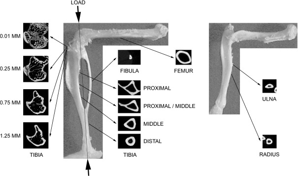
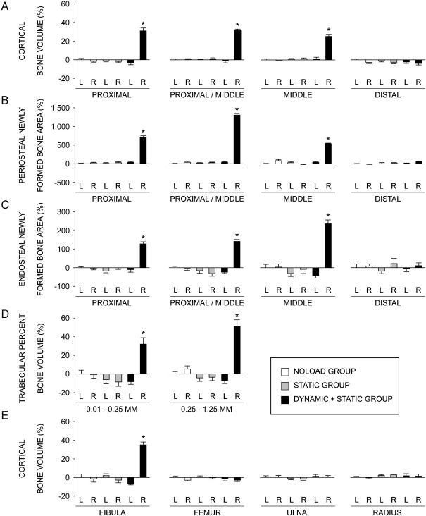
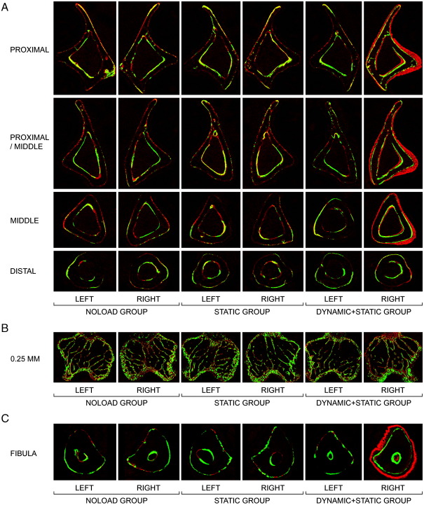
In order to validate whether bones' functional adaptation to mechanical loading is a local phenomenon, we randomly assigned 21 female C57BL/6 mice at 19 weeks of age to one of three equal numbered groups. All groups were treated with isoflurane anesthesia three times a week for 2 weeks (approximately 7 min/day). During each anaesthetic period, the right tibiae/fibulae in the DYNAMIC+STATIC group were subjected to a peak dynamic load of 11.5 N (40 cycles with 10-s intervals between cycles) superimposed upon a static "pre-load" of 2.0 N. This total load of 13.5 N engendered peak longitudinal strains of approximately 1400 microstrain on the medial surface of the tibia at a middle/proximal site. The right tibiae/fibulae in the STATIC group received the static "pre-load" alone while the NOLOAD group received no artificial loading. After 2 weeks, the animals were sacrificed and both tibiae, fibulae, femora, ulnae and radii analyzed by three-dimensional high-resolution (5 mum) micro-computed tomography (microCT). In the DYNAMIC+STATIC group, the proximal trabecular percent bone volume and cortical bone volume at the proximal and middle levels of the right tibiae as well as the cortical bone volume at the middle level of the right fibulae were markedly greater than the left. In contrast, the left bones in the DYNAMIC+STATIC group showed no differences compared to the left or right bones in the NOLOAD or STATIC group. These microCT data were confirmed by two-dimensional examination of fluorochrome labels in bone sections which showed the predominantly woven nature of the new bone formed in the loaded bones. We conclude that the adaptive response in both cortical and trabecular regions of bones subjected to short periods of dynamic loading, even when this response is sufficiently vigorous to stimulate woven bone formation, is confined to the loaded bones and does not involve changes in other bones that are adjacent, contra-lateral or remote to them.
(c) 2009 Elsevier Inc. All rights reserved.
Figures
References
-
- Frost H.M. Bone “mass” and the “mechanostat”: a proposal. Anat. Rec. 1987;219:1–9. - PubMed
-
- Torrance A.G., Mosley J.R., Suswillo R.F., Lanyon L.E. Noninvasive loading of the rat ulna in vivo induces a strain-related modeling response uncomplicated by trauma or periostal pressure. Calcif. Tissue Int. 1994;54:241–247. - PubMed
-
- Raab-Cullen D.M., Akhter M.P., Kimmel D.B., Recker R.R. Periosteal bone formation stimulated by externally induced bending strains. J. Bone Miner. Res. 1994;9:1143–1152. - PubMed
-
- Turner C.H., Forwood M.R., Rho J.Y., Yoshikawa T. Mechanical loading thresholds for lamellar and woven bone formation. J. Bone Miner. Res. 1994;9:87–97. - PubMed
-
- Akhter M.P., Cullen D.M., Pedersen E.A., Kimmel D.B., Recker R.R. Bone response to in vivo mechanical loading in two breeds of mice. Calcif. Tissue Int. 1998;63:442–449. - PubMed
Publication types
MeSH terms
Substances
Grants and funding
LinkOut - more resources
Full Text Sources
