. 2009 Sep;124(3):496-506, 506.e1-6.
doi: 10.1016/j.jaci.2009.06.046.

Filaggrin deficiency confers a paracellular barrier abnormality that reduces inflammatory thresholds to irritants and haptens

Affiliations

Filaggrin deficiency confers a paracellular barrier abnormality that reduces inflammatory thresholds to irritants and haptens

Tiffany C Scharschmidt et al. J Allergy Clin Immunol. 2009 Sep.

Abstract

Background: Mutations in the human filaggrin gene (FLG) are associated with atopic dermatitis (AD) and are presumed to provoke a barrier abnormality. Yet additional acquired stressors might be necessary because the same mutations can result in a noninflammatory disorder, ichthyosis vulgaris.

Objective: We examined here whether FLG deficiency alone suffices to produce a barrier abnormality, the basis for the putative abnormality, and its proinflammatory consequences.

Methods: By using the flaky-tail mouse, which lacks processed murine filaggrin because of a frameshift mutation in the gene encoding profilaggrin that mimics some mutations in human AD, we assessed whether FLG deficiency provokes a barrier abnormality, further localized the defect, identified its subcellular basis, and assessed thresholds to irritant- and hapten-induced dermatitis.

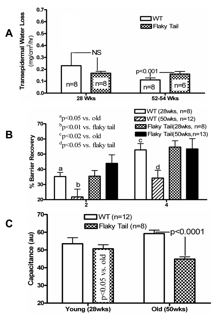
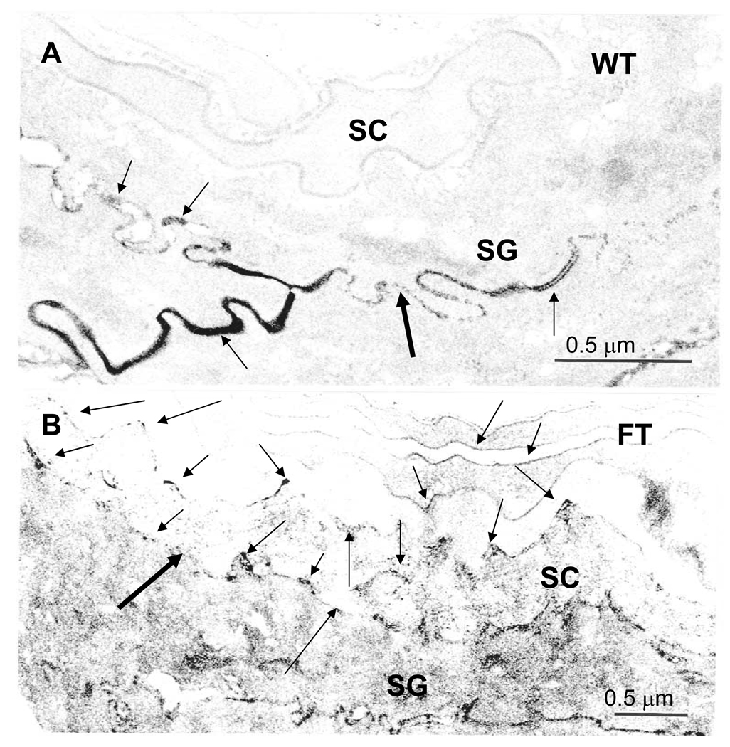
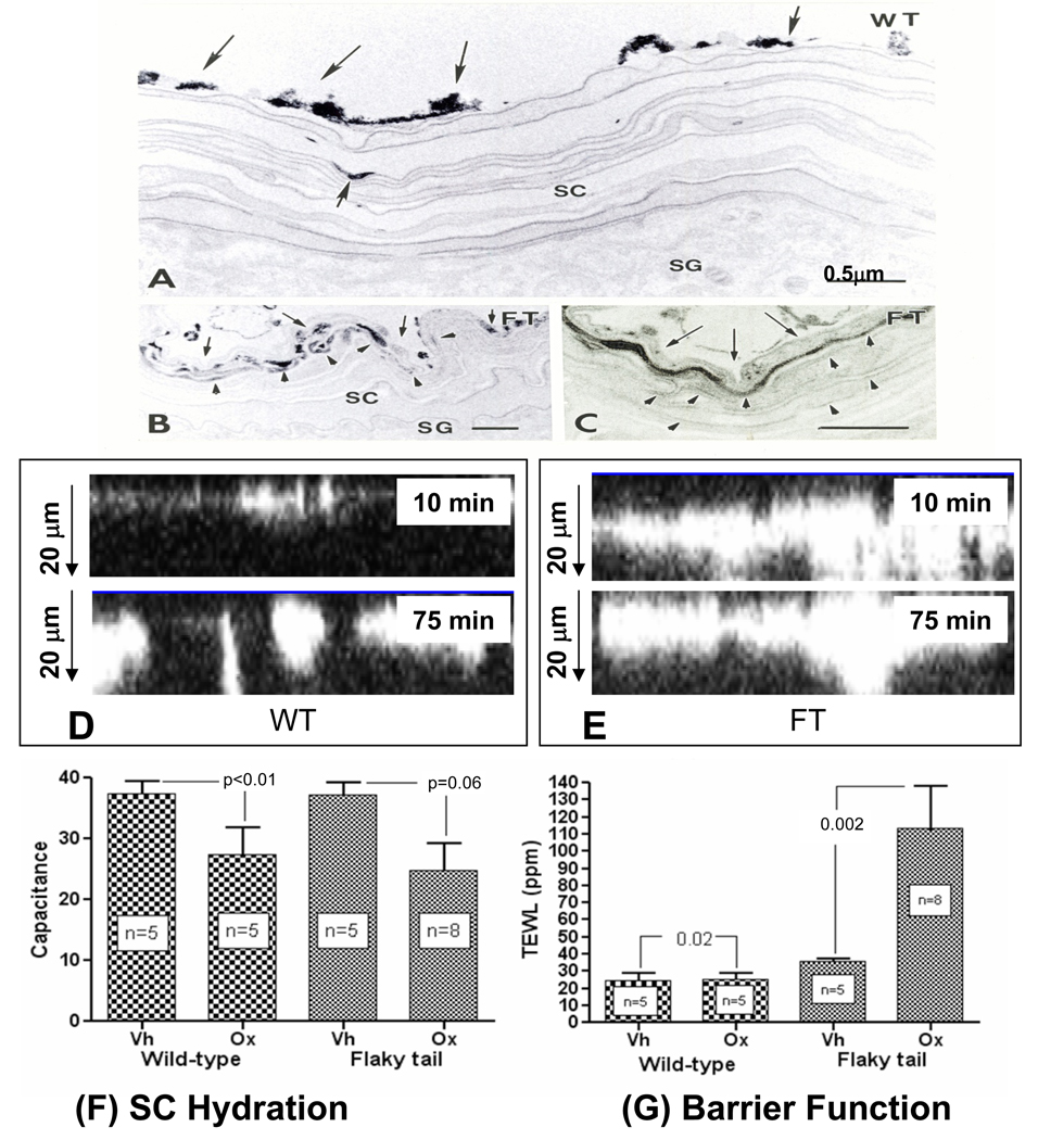
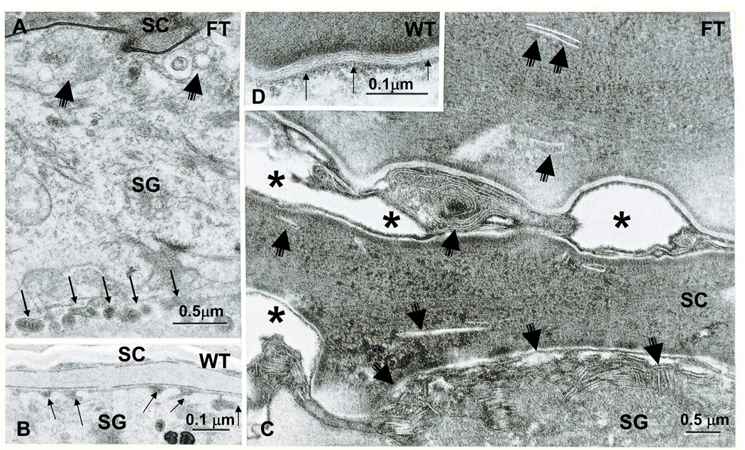
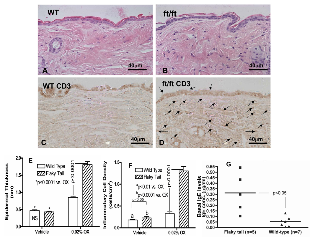
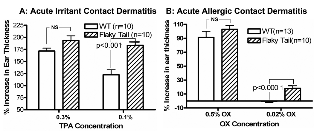
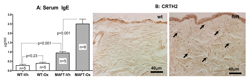
Results: Flaky-tail mice exhibit low-grade inflammation with increased bidirectional, paracellular permeability of water-soluble xenobiotes caused by impaired lamellar body secretion and altered stratum corneum extracellular membranes. This barrier abnormality correlates with reduced inflammatory thresholds to both topical irritants and haptens. Moreover, when exposed repeatedly to topical haptens at doses that produce no inflammation in wild-type mice, flaky-tail mice experience a severe AD-like dermatosis with a further deterioration in barrier function and features of a T(H)2 immunophenotype (increased CRTH levels plus inflammation, increased serum IgE levels, and reduced antimicrobial peptide [mBD3] expression).

Conclusions: FLG deficiency alone provokes a paracellular barrier abnormality in mice that reduces inflammatory thresholds to topical irritants/haptens, likely accounting for enhanced antigen penetration in FLG-associated AD.


Figures

Figure 1
Figure 1. Abnormal Barrier Function and Stratum Corneum Hydration in Flaky Tail Mice
A: Basal transepidermal water loss (TEWL) was assessed with an electrolytic analyzer as mg/cm2/hr, and expressed as (mean) ± SEM. B: Barrier disruption was achieved with repeated tape stripping until TEWL levels ≥ 3× normal; then % recovery from an initial ‘0’ value, was measured 2 and 4 hrs after stripping, and expressed as mean +/−SEM. C: Stratum corneum hydration was measured as electrical capacitance on shaved back skin in flaky tail and wild-type mice, and shown as changes in arbitrary units (AU) +/− SEM (n=8–13, as indicated).
Figure 2
Figure 2. Abnormal Paracellular Permeability Barrier to a Subcutaneous, Water-Soluble Tracer in Flaky Tail Mice
Explants of back skin from sex-matched, 12 wk old, wild-type (WT) and flaky tail (FT) mice (n = 3 each) were immersed dermis-side-downward on a solution containing 4% colloidal lanthanum nitrate (pH 7.5) for 30 min to 2 hr, followed by fixation in Karnovsky’s solution (see Methods). Colloidal lanthanum travels outward through the extracellular spaces, but does not reach the stratum corneum (SC) in WT skin (A, arrows). In contrast, in FT skin, lanthanum tracer extends into the lower SC, primarily via the extracellular spaces (B, arrows), suggesting impaired ‘inside-to-outside’ barrier function. SG = stratum granulosum; A+B, osmium tetroxide post-fixation; Mag bars = 0.5 µm.
Figure 3
Figure 3. Epicutaneous Tracer Penetrates Flaky Tail Stratum Corneum via the Extracellular Spaces
A–C: Flanks of flaky tail (FT) and wild-type (WT) mice (n = 3 each) were immersed in 4% lanthanum nitrate solution for 30 min to 2 hrs, followed by aldehyde fixation. After 2 hrs, little or no tracer entered the stratum corneum (SC) of WT mice (A, single small arrow). In contrast, abundant tracer reaches ≥4 layers into the SC in FT mice (B, arrowheads), which on higher magnification [C] can be seen to localize to extracellular domains (C, arrowheads). D+E: Calcium green was applied to freshly-obtained explants from ft/ft and wt mice (n = 3 each), and penetration was assessed by dual-photon confocal microscopy, along the ‘z’ axis after 10 and 75 min. Note much deeper ‘outside-to-inside’ penetration of calcium green in ft/ft mice at both time points. Flaky tail (FT) mice, repeatedly-challenged with low-dose Ox, display a more severe barrier abnormality (G), but no further decline in stratum corneum (SC) hydration in comparison to wild-type (WT) mice (F). A–C: Osmium tetroxide post-fixation; mag bar = 0.5 µm.
Figure 4
Figure 4. Abnormalities in the Lamellar Body Secretory System in Young Flaky Tail Mice
Partial failure of lamellar body exocytosis is evident in flaky tail (FT) epidermis (A, C). Note lamellar bodies lined up in peripheral cytosol (A, multiple thin arrows); decreased secreted material at stratum granulosum (SG)-stratum corneum (SC) interface (A&C, short, fat arrows); decreased numbers of lamellar bilayers (C): delayed maturation of lamellar bilayers (C); and entombed lamellar body contents within the corneocyte cytosol (C, short, thin arrows). B&D: Normal lamellar body secretion (B, arrows) and extracellular lamellar bilayers in wild-type (WT) epidermis (D, thin arrows) A&B, osmium tetroxide post-fixation; C&D, ruthenium tetroxide post-fixation. Mag bars = A&C, 0.5 µm; B&D, 0.1 µm.
Figure 5
Figure 5. Reduced Threshold for Development of AD-Like Inflammation in Response to Repeated Ox Challenges in Flaky Tail Mice
Flaky tail (FT) and wild-type (WT) mice were sensitized to Ox, and then repeatedly challenged with a subthreshold concentration of Ox (0.02%) every other day to a shaved area of back skin for a total of 10 challenges. Under basal conditions, FT mice display low-grade inflammation (F, see also suppl. Fig. 3C vs. A). FT skin displayed much more prominent erythema, scaling and excoriations than does WT mouse skin (see text), a change that is associated with a modest inflammatory infiltrate (B vs. A & suppl. Fig. 3C), that was enriched in CD3+ dermal lymphocytes (D vs. C). Quantitative changes in epidermal hyperplasia and inflammation are shown in E&F. G: Serum IgE levels are significantly elevated in most flaky tail mice, even under basal conditions. Mag bars = 40 µm.
Figure 6
Figure 6. Altered Sensitivity to Irritant/Hapten-Induced Allergic Contact Dermatitis in Flaky Tail Mice
In young, wild-type mice, TPA induces irritant contact dermatitis, while Ox induces allergic contact dermatitis following a single, full-strength challenge dose . While higher concentrations produced comparable changes in young wide-type (WT) and flaky tail mice, lower doses of either TPA (A) or Ox (B) induce inflammation only in flaky tail mice. Changes in ear thickness correlate with severity of inflammation (see suppl Fig. 4).
Figure 7
Figure 7. Flaky Tail Mice Demonstrate Increased Th2 Response to Repeated Low-Dose Ox Challenges
Serum was collected from young flaky tail (FT) and wild-type (WT) mice before and after Ox sensitization, and either ten low-dose Ox (0.02%) or vehicle treatments, followed by assessment of IgE levels by ELISA. While no difference from baseline was seen between vehicle and Ox-treated WT mice, a large increase was observed in FT mice (B). Likewise, low-dose Ox-challenged FT mice demonstrate increased staining for CRTH2-bearing cells (A, arrows) in Ox-treated, FT mice. Mag bars = 40 µm.

References

    1. Elias PM, Hatano Y, Williams ML. Basis for the barrier abnormality in atopic dermatitis: outside-inside-outside pathogenic mechanisms. J Allergy Clin Immunol. 2008;121:1337–1343. - PMC - PubMed
    1. Elias PM, Steinhoff M. "Outside-to-inside" (and now back to "outside") pathogenic mechanisms in atopic dermatitis. J Invest Dermatol. 2008;128:1067–1070. - PMC - PubMed
    1. Cork MJ, Robinson DA, Vasilopoulos Y, Ferguson A, Moustafa M, MacGowan A, et al. New perspectives on epidermal barrier dysfunction in atopic dermatitis: gene-environment interactions. J Allergy Clin Immunol. 2006;118:3–21. quiz 2–3. - PubMed
    1. Nemoto-Hasebe I, Akiyama M, Nomura T, Sandilands A, McLean WH, Shimizu H. Clinical severity correlates with impaired barrier in filaggrin-related eczema. J Invest Dermatol. 2009;129:682–689. - PubMed
    1. Boguniewicz M, Leung DY. 10. Atopic dermatitis. J Allergy Clin Immunol. 2006;117:S475–S480. - PubMed

Publication types